Download
1 / 41
410 likes | 549 Vues
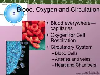
The heart and circulation Transportation- oxygen and carbon dioxide red blood cells Regulation-hormones, temperature Protection- against blood loss (clotting); infection (immune system). Components of the circulatory system Cardiovascular system- heart and blood vessels

E N D
The heart and circulation Transportation- oxygen and carbon dioxide red blood cells Regulation-hormones, temperature Protection- against blood loss (clotting); infection (immune system)
Components of the circulatory system Cardiovascular system- heart and blood vessels Lymphatic system- lymph nodes, lymphatic vessels
Plasma- water, ions, proteins Plasma proteins albumin- provides osmotic pressure globulins- alpha and beta- transport gamma- antibodies (produced by lymphocytes; other proteins by liver) fibrinogen- clotting Plasma volume regulated by hormones like ADH
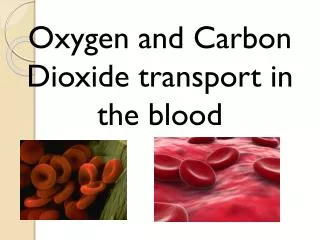
“Formed elements” (cells) Erythrocytes- (red blood cells) no nuclei or mitochondria circulate for about 120 days 280 million hemoglobin molecules per cell Leukocytes (white blood cells) granular and agranular granular: neutrophils, eosinophils, basophils agranular: lymphocytes, monocytes capable of amoeboid movement Platelets- involved in clotting
Hematopoiesis- blood cell formation Erythropoiesis- red blood cell formation Leukopoiesis- leukocyte formation Controlled by cytokines that regulate cell growth and formation Growth factors Colony-stimulating factors Erythropoietin
Red blood cell antigens and blood typing Antigen: a molecule that is recognized as foreign by the immune system Lots of these: several different types of antigens found on red blood cells (RBCs) ABO system especially important Four blood types: A, B, AB, O A and B are dominant, O is recessive
People with type A blood can tolerate type A blood from other individuals But type A people make antibodies to type B antigens People with type AB can tolerate all blood types: universal recipient (of CELLS) People with type O blood can donate to all but have antibodies to both A and B antigens: universal donor (of CELLS)
Rh antigen is another important blood group antigen- people are either Rh-positive or Rh-negative Significance: when Rh-negative woman is pregnant with Rh-positive baby Mother might produce antibodies to Rh antigen on fetus’ blood cellserythroblastosis fetalis, or hemolytic disease of the newborn Prevention: Rhogam
Blood clotting- hemostasis Vasoconstriction Formation of platelet plug platelets stick to collagen proteins coated by von Willebrand factor (secreted by damaged endothelium) platelets stick together and secrete factors that promote vasoconstriction- cascade Formation of fibrin web that surrounds plug intrinsic pathway- “spontaneous” extrinsic- initiated by factors from damaged tissue
Clot dissolution Factor XIIkallikreinplasminogenplasmin Anticoagulants calcium chelators heparin- inactivates thrombin coumarin- inhibits vitamin K activation aspirin- inhibits prostaglandin production, affects platelet release reaction
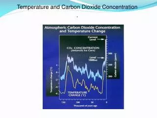
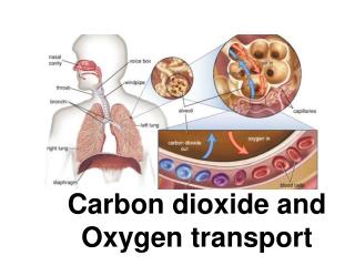
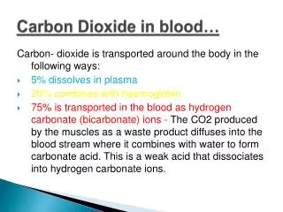
Acid-base balance of blood Blood plasma pH ranges between 7.35 and 7.45 CO2 + H2OH2CO3H++HCO3- Carbon dioxide transported through blood concentration controlled by breathing Kidneys release excess H+ and reabsorb bicarbonate pH too low- acidosis; pH too high- alkalosis metabolic ore respiratory
Acidosis respiratory- hypoventilation metabolic- excess production of ketone bodies, fatty acids, lactic acid diabetes- fat metabolism diarrhea- loss of bicarbonate
Alkalosis respiratory- hyperventilation metabolic- too much bicarbonate excess vomiting loss of fatty acids, ketone bodies, etc. loss of acids in gastric juice So critical blood chemicals are carbon dioxide and bicarbonate
Structure of the heart Two atria, two ventricles Atria receive blood from venous system Ventricles pump blood into arterial system Septum separates right from left side
Valves embedded in fibrous skeleton AV valve between right atrium and ventricle- tricuspid valve AV valve between left atrium and ventricle bicuspid (mitral) valve Semilunar valves at base of pulmonary artery and aorta
Cardiac cycle and heart sounds Contraction- systole Relaxation- diastole Atria contract simultaneously Then ventricles contract (while atria are relaxed) Stroke volume- amount of blood ejected from ventricles during systole end-systolic volume- what’s left
Electrical activity of the heart Myocardial cells beat automatically Action potential is usually originated in sinoatrial node Spontaneous depolarization (pacemaker potential) diffusion of calcium through slow channels threshold- fast calcium channels open, voltage regulated sodium channels open repolarization produced through diffusion of potassium
Other parts of the heart can produce pacemaker potentials Depolarize more slowly than SA node; usually stimulated by action potentials from SA node before they could start their own pacemaker potentials “ectopic pacemakers” can set a rhythm if SA node conduction is blocked; pace will be slower
Heart muscle cannot sustain contraction Long refractory periods- heart cannot be stimulated until it has relaxed from previous contraction Arrhythmias- something affects the cardiac cycle; treatment depends on what it is Fast Na channel blockers Slow Ca channel blockers -adrenergic receptor blockers
Electrocardiogram Conduction of electrical potentials through heart P wave- atrial depolarization QRS- ventricular depolarization beginning of systole T wave- repolarization of the ventricles beginning of diastole
Blood vessels- arteries and veins Arteries, arterioles, capillaries Veins and venules Arteries are more muscular Veins have valves
Capillaries deliver blood to cells Specialized types of capillaries in different organs Fenestrated- kidneys, endocrine glands, intestines Discontinuous- bone marrow, liver and spleen Continuous- everywhere else
Veins Veins can expand to accommodate increasing amounts of blood; arteries can’t Venous pressure is low compared to arterial pressure Blood flow through veins is facilitated by: contraction of skeletal muscles valves that prevent backflow
Atherosclerosis Damage to endothelium “Fatty streaks” (macrophages and lymphocytes) Fibrous plaques High blood cholesterol, LDL contribute to atherosclerosis HDL also help transport cholesterol, but do not contribute to atherosclerosis
Arrhythmias Bradycardia- slow rate (less than 60 bpm) Tachycardia- fast rate (more than 100 bpm) Can occur normally; is abnormal if rate increases during rest (ectopic pacemakers) Flutters- extremely rapid contractions Fibrillation- different groups of fibers are activated so coordinated pumping of chambers is not possible
Lymphatic system Fluid transport from tissues Fat transport from intestines Immune response