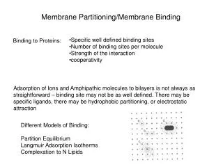
Membrane Dynamics
Membrane Dynamics. 5. About this Chapter. Mass balance and homeostasis Diffusion Protein-mediated, vesicular, and transepithelial transport Osmosis and tonicity The resting membrane potential Insulin secretion. Mass Balance in the Body. Figure 5-2. Mass Balance and Homeostasis.

Membrane Dynamics
E N D
Presentation Transcript
About this Chapter • Mass balance and homeostasis • Diffusion • Protein-mediated, vesicular, and transepithelial transport • Osmosis and tonicity • The resting membrane potential • Insulin secretion
Mass Balance in the Body Figure 5-2
Mass Balance and Homeostasis • Clearance • Rate at which a molecule disappears from the body • Mass flow = concentration volume flow • Homeostasis equilibrium • Osmotic equilibrium • Chemical disequilibrium • Electrical disequilibrium
Homeostasis Distribution of solutes in the body fluid compartments The compartments in the body are in a state of chemical disequilibrium Figure 5-3a
Homeostasis Figure 5-3b
Diffusion Map of membrane transport Membranes are selectively permeable Figure 5-4
Diffusion: Seven Proprieties • Passive process • High concentration to low concentration • Net movement until concentration is equal • Rapid over short distances • Directly related to temperature • Inversely related to molecular size • In open system or across a partition
Simple Diffusion Fick’s law of diffusion Figure 5-6
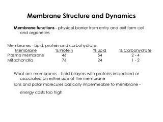
Membrane Proteins Function • Structural proteins • Enzymes • Membrane receptor proteins • Transporters • Channel proteins • Carrier proteins
Membrane Transport Proteins Water channels and ion channels are examples of open channels Figure 5-9a
Membrane Transport Proteins Figure 5-9b
Gating of Channel Proteins Gated channels are either chemically gated or voltage-gated channels Figure 5-11
Types of Carrier-Mediated Transport Figure 5-12a
Types of Carrier-Mediated Transport Figure 5-12b
Types of Carrier-Mediated Transport Carrier proteins never create a continuous passageway Figure 5-12c
Facilitated Diffusion Diffusion of glucose into cell • How is the concentration gradient maintained for glucose? Figure 5-15
Primary Active Transport ECF 1 ATP ADP 5 2 3 Na+ from ICF bind ICF P ATPase is phosphorylated with Pi from ATP. 2 K+ released into ICF Protein changes conformation. Protein changes conformation. P 3 K+ released into ICF 4 3 2 K+ from ECF bind P P Mechanism of the Na+-K+-ATPase ATP is used as an energy source Figure 5-17
Primary Active Transport ECF 1 3 Na+ from ICF bind ICF Figure 5-17, step 1
Primary Active Transport ECF 1 ATP ADP 2 3 Na+ from ICF bind ICF P ATPase is phosphorylated with Pi from ATP. Figure 5-17, steps 1–2
Primary Active Transport ECF 1 ATP ADP 2 3 Na+ from ICF bind ICF P ATPase is phosphorylated with Pi from ATP. Protein changes conformation. 3 K+ released into ICF 3 P Figure 5-17, steps 1–3
Primary Active Transport ECF 1 ATP ADP 2 3 Na+ from ICF bind ICF P ATPase is phosphorylated with Pi from ATP. Protein changes conformation. 3 K+ released into ICF 4 3 2 K+ from ECF bind P P Figure 5-17, steps 1–4
Primary Active Transport ECF 1 ATP ADP 5 2 3 Na+ from ICF bind ICF P ATPase is phosphorylated with Pi from ATP. 2 K+ released into ICF Protein changes conformation. Protein changes conformation. P 3 K+ released into ICF 4 3 2 K+ from ECF bind P P Figure 5-17, steps 1–5
Secondary Active Transport 1 Na+ binds to carrier. 3 Glucose binding changes carrier conformation. Intracellular fluid Lumen of intestine or kidney Na+ Na+ SGLT protein Glu Glu [Na+] high [Glucose] low [Na+] low [Glucose] high Na+ released into cytosol. Glucose follows. 4 2 Na+ binding creates a site for glucose. Na+ Na+ Glu Glu Mechanism of the SGLT Transporter Figure 5-18
Secondary Active Transport 1 Na+ binds to carrier. Intracellular fluid Lumen of intestine or kidney Na+ SGLT protein Glu [Na+] high [Glucose] low [Na+] low [Glucose] high Figure 5-18, step 1
Secondary Active Transport 1 Na+ binds to carrier. Intracellular fluid Lumen of intestine or kidney Na+ SGLT protein Glu [Na+] high [Glucose] low [Na+] low [Glucose] high 2 Na+ binding creates a site for glucose. Na+ Glu Figure 5-18, steps 1–2
Secondary Active Transport 1 Na+ binds to carrier. 3 Glucose binding changes carrier conformation. Intracellular fluid Lumen of intestine or kidney Na+ Na+ SGLT protein Glu Glu [Na+] high [Glucose] low [Na+] low [Glucose] high 2 Na+ binding creates a site for glucose. Na+ Glu Uses the energy of one molecule moving down its concentration gradient Figure 5-18, steps 1–3
Secondary Active Transport 1 Na+ binds to carrier. 3 Glucose binding changes carrier conformation. Intracellular fluid Lumen of intestine or kidney Na+ Na+ SGLT protein Glu Glu [Na+] high [Glucose] low [Na+] low [Glucose] high Na+ released into cytosol. Glucose follows. 4 2 Na+ binding creates a site for glucose. Na+ Na+ Glu Glu Figure 5-18, steps 1–4