Download
1 / 35
350 likes | 606 Vues
Membrane Structure and Dynamics. CH353 February 14, 2008. Summary. Membrane Lipids Diversity and distribution Biophysics: phase transitions and diffusion Structures Membrane Proteins Classification Structures Functions: Membrane shape Membrane fusion Cell adhesion.

E N D
Membrane Structure and Dynamics CH353 February 14, 2008
Summary Membrane Lipids • Diversity and distribution • Biophysics: phase transitions and diffusion • Structures Membrane Proteins • Classification • Structures • Functions: • Membrane shape • Membrane fusion • Cell adhesion
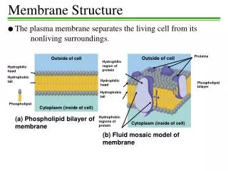
General Properties of Biomembranes • Non-covalent assembly of lipid and protein – fluid mosaic • Lipids spontaneously form a bilayer 5–8 nm thick • Hydrophobic interior with hydrophillic surfaces • Selectively impermeable to polar molecules • Creates a barrier for separating aqueous environments • Movement of lipids and proteins • Rapid diffusion within each monolayer • Slow diffusion from one monolayer to another • Structure and function depends on both lipid and protein • Asymmetry of lipids and proteins in each monolayer • Electrochemical differences across membrane
Diversity of Lipid Components Factors determining fluidity, thickness, shape and activity of biomembrane Type of Lipid • glycerolipid, sphingolipid, cholesterol Variations in acyl or ether groups • length, unsaturation Head Group Alcohol • choline, ethanolamine, inositol, serine, carbohydrate >1000 different combinations of acyl and head groups per eukaryotic cell Sprong et al. 2001, Nature Rev. Mol. Biol. 2: 504.
Distribution of Lipids in Organelles • Cholesterol in plasma membrane • Cardiolipin in inner mitochondrial • Sphingolipids in lysosomal
Distribution of Lipids in Bilayer Erythrocyte plasma membrane • Inside (anionic groups) • phosphatidylethanolamine • phosphatidylserine • phosphatidylinositols • phosphatidic acid • Outside (neutral groups) • phosphatidylcholine • sphingomyelin • Both • cholesterol
Fluidity of Biomembranes • Pure lipids have a phase transition: gel ↔ fluid paracrystalline↔ liquid-disordered • Biomembranes having mixtures of lipids exist in liquid-ordered state • Cell changes the composition of its lipid bilayer to maintain that state • Less fluid membranes have longer and more saturated acyl groups • cis double bonds disrupt packing • sterols pack with saturated acyl groups; both ordered and fluid
Phase Diagram determined by EPR (Electron Paramagnetic Resonance) ld 40 ld so 30 lo Temperature (ºC) 20 lo so 10 0 0.0 0.1 0.2 0.3 Cholesterol : Lecithin Effect of Cholesterol on Membranes Meer et al. 2008, Nature Rev. Mol. Biol. 9: 112.
Lateral Movement of Lipids • Rapid diffusion of lipids within the monolayer • Motion is restricted by cell structures • Organelles • Cell-cell junctions • Cytoskeletal elements • Fluorescence microscopy of single lipid • Rapid diffusion within a region with jumps to other regions
Lateral Movement of Lipids • Fluorescence Recovery After Photobleaching (FRAP) Analysis • Outer leaflet of membrane is labeled with probe • Laser bleaches a spot on labeled lipids • Fluorescence microscopy shows rapid lateral motion of lipids into the bleached spot on membrane
Transverse Movement of Lipids • Movement of lipids from one bilayer to another is relatively slow • Biological flipping of lipids is catalyzed with proteins • P4 ATPases • ATP binding cassette (ABC) transporters
Movement of Lipids from Monolayer • t1/2 of spontaneous transfer for various lipids • loss of head group alcohol ↑ transbilayer diffusion • loss of fatty acyl group ↑ interbilayer transfer • liquid-ordered domains ↓ both types of transfer • cholesterol shows rapid transbilayer diffusion Hothuis & Levine 2005, Nature Rev. Mol. Biol. 6: 209.
Cross-Sectional Shapes of Lipids • amphipathic molecules with a single hydrocarbon chain form micelles • detergents • fatty acids • lysoglycerophospholipids • some amphipathic molecules with two hydrocarbon chains form bilayers • phosphatidylcholine • some lipids do not form stable bilayers • cholesterol • phosphatidylethanolamine
Shapes of Lipids Determine Structures • Lysophosphatidylcholine (lysolecithin) – conical • forms micelles • Phosphatidylcholine (lecithin) • cylindrical • forms lipid bilayer • Phosphatidylethanolamine • inverted conical • forms inverse micelles (inverted hexagonal phase) • Lipids with non-cylindricalcross sections may have special cellular functions Sprong et al. 2001, Nature Rev. Mol. Biol. 2: 504.
Lipids Determine Membrane Thickness • Phosphatidylcholine bilayer: • 3.5 nm thick • Phosphatidylcholine + cholesterol bilayer: • 4.0 nm thick • Sphingomyelin + cholesterol bilayer: • 4.7 nm thick • Lipid rafts – local regions of thicker membrane with more sphingolipid and cholesterol Sprong et al. 2001, Nature Rev. Mol. Biol. 2: 504.
Types of Membrane Proteins • Integral Proteins • Covalently attached to lipid or embedded in membrane • Require extraction with agents that interfere with hydrophobic interactions, e.g. detergents • Peripheral Proteins • Non-covalent interactions with integral proteins or lipids • Can be removed using mild methods disrupting ionic interactions and H-bonds
Types of Integral Membrane Proteins Type I – one transmembrane helix, N-term outside Type II – one transmembrane helix, C-term outside Type III – multiple transmembrane helices on single polypeptide Type IV – multiple transmembrane helices on separate polypeptides Type V – proteins covalently bound to lipid Type VI – proteins with covalently bound lipid and transmembrane helix
Glycophorin A type I membrane protein • Amino-terminal domain has polar amino acids and is glycosylated • 15 O-linked tetrasaccharides • 1 N-linked glycan • Transmembrane domain has hydrophobic amino acids • Carboxy-terminal domain has polar amino acids
Bacteriorhodopsin A type III membrane protein • 7 transmembrane α-helices • Each helix is composed of hydrophobic amino acids • Loops joining helices have polar amino acids • A light-driven H+ pump • Analogous to rhodopsin (a G protein-coupled receptor)
Prediction of Transmembrane α-Helices • Each amino acid is given a hydropathy index based on free energy of transfer to water • Hydropathy of peptides within an amino acid sequence are calculated and plotted vs residue number • Transmembrane peptides are ones with hydropathy > 0 for 20–25 amino acids
Membrane Proteins with β-Barrel Structures • Bacterial β-barrel porins • Transmembrane β-sheets have alternating polar and non-polar amino acids • Cannot use scanning hydropathy method for predicting membrane spanning β-sheets
Lipid-Linked Membrane Proteins • Integral proteins can be attached to membrane by covalently bound lipid • Acylated • palmitoyl on Cys or Ser • N-terminal myristoyl • Prenylated • C-terminal farnesyl or geranylgeranyl group • GPI anchor • C-terminal glycosyl phosphatidylinositol
Imaging Membrane Ultrastructure Using Electron Microscopy • Freeze-fracture/freeze-etch scanning EM • Specimen rapidly frozen then fractured along plane parallel to lipid bilayers • Sample is then shadowed with platinum and organic material is dissolved • The freeze-etched metal replica is then analyzed by scanning EM Image of thylakoid membrane
Imaging Membrane Ultrastructure Using Atomic Force Microscopy
Structure of Membrane Rafts • Microdomains on surface of plasma membrane • Sphingolipid and cholesterol in liquid-ordered state • Surrounding lipid in bilayer in liquid-disordered state • Resistant to solubilization • Segregate proteins based on attached lipid (fatty acyl groups pack better) • Caveolin embedded in some (inward curvature)
Structure of Membrane Caveolae • Caveolae “little caves” caused by the binding of numerous caveolins in membrane rafts • Both bilayers are involved in the rafts • May have important functions in membrane trafficking and signal transduction
Cell Adhesion Proteins • Cadherins • homophilic interactions • Immunoglobulin-like domains • N-CAM (homophilic) • I-CAM (bind integrin) • Selectins • bind carbohydrate • Integrins • Combinations of α and β • heterophilic interactions with various ligands • 2-way signal tranduction (integrin activation)
Cell Adhesion Molecules Participate in Leukocyte Activation and Extravasation • Inflammation causes presentation of P-selectin and platelet activating factor • P-selectin interacts transiently with carbohydrate ligands on leukocytes • PAF activates leukocyte causing activation of integrin (inside-out signalling) • Integrins adhere firmly with ICAM-1 and ICAM-2 on endothelial cells
Integrin Heterodimeric Complexes natural combinations of integrin α and β subunits and their ligands
SNARE Conformational Cycle • SNARE complexes: 4 parallel α helices each C-term in membrane • Ca2+ required for binding R-SNARE with Q-SNAREs (acceptor) • ATP needed for dissociation of SNARE complexes Jahn & Scheller 2006, Nature Rev. Mol. Biol. 7: 631
Distinct SNARE Combinations for Different Membrane Fusions Jahn & Scheller 2006, Nature Rev. Mol. Biol. 7: 631