Download
1 / 47
470 likes | 737 Vues
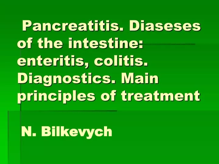
Pancreatitis . Diaseses of the intestine : enteritis , colitis . Diagnostics . Main principles of treatment N. Bilkevych. The pancreas is an elongated, tapered gland that is located behind the stomach and secretes digestive enzymes and the hormones insulin and glucagon.

E N D
Pancreatitis. Diaseses of the intestine: enteritis, colitis. Diagnostics. Main principles of treatmentN. Bilkevych
The pancreas is an elongated, tapered gland that is located behind the stomach and secretes digestive enzymes and the hormones insulin and glucagon.
The pancreas is located posterior to the abdomen. It contains cells that secrete the hormone insulin, and cells that secrete digestive enzymes that aid in the breakdown of food in the gastrointestinal tract. The pancreas secretes these enzymes into the pancreatic duct, which joins the common bile duct from the liver and drains into the small intestine.
Chronic pancreatitis (CP) - • Chronic inflammatory affection of pancreatic gland parenchymawhich lasts over 6 month з with exocrinic parenchymal destruction, fibrosisand on later stages - endocrinic parenchymal destruction.
Ethiology Secondary pancreatitis : diseases of bile ducts (30-40 %) diseases of a duodenum pathology of duodenal papilla:primary (tumor,papilitis) and secondary (oddy’s sphincter dyskinesia, skars) diseases of a liver diseases of intestine viralinfections (epidemic parotitis) allergy hyperlipidemia hyperparathyreosis traumas Primary pancreatitis: • Alсohol abuse (70-80%) • Systematic fat food intake • Medicaments(azathioprin, izoniazid, tetracycline, sulfa drugs) • Protein defficiency (kwashiorkor) • Hereditary • Ischemic (affection of pancreaticvessels) • Idiopatic
Clinical classification According to functional characteristics 1. With exocrinic functional disorders. 2. With endocrinic disorders. Phase of the disease: -exacerbation, -remission. Complications
Clinical pattern Leading syndromes: • Pain • Dyspepsy • Exocrinic secratory disfunction and syndromes of maldigestia and malabsorption with progressive body weight loss • Exocrinic secratory disfunction (pancreatic diabetes mellitus) • Asthenic syndrome
Clinical pattern Pain syndrome Dejarden’s point
Dyspeptic syndrome Appetite is decreased or absent, hypersalivation, nausea, Vomiting without benefition, meteotism, Disordered stool (prevalence of diarrhoea or change of diarrhoea and constipation). Clinical pattern
Clinical pattern Syndrome of exogenous dysfunction - • -“pancreatic" diarrhoea is characterised with large volume(polyfecalia), of greish color, with unpleasant smell and fatty (steatorrhoea). • body mass loss with frequent development of osteoporosis (painin bones) because of calcium loss andvitamin D deficiency.
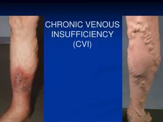
Spider angiomata Red spots on the skin of the chest, abdomen and back. They are round, don’t disappear after pressing in them (microaneurisms) Clinical pattern
Laboratory diagnostics: 1. Complete blood count: in 25 % – leucocytosis, increased ESR. 2. Assessment of pancreatic enzymes in the blood (amylase, lipase, tripsine in blood and urine — increased in exacerbation). !!!increased level of α-amylaseon 1-st day of the diseasein 85-90 % of cases, on2-nt day – in 60-70 %, on3-rd day – in 40-50 %.
Instrumental diagnostics • computer tomography:diagnostic information similliar toultrasound examination,is executet in suspition on cancer or cysts of the gland; • MRT: visualization of normal and pathologically changed pancreatic ducts, stones;
Endoscopic examination • An endoscope, with a camera on its end, is passed down the esophagus, through the stomach, and into the small intestine. The entrance of the pancreatic duct into the small intestine can be viewed through the endoscope. A special instrument on the end of the endoscope can then be passed into the pancreatic duct and the gallstone is extracted. Very rarely pancreatitis is severe enough to require surgery, which is usually performed when the pancreas becomes infected. Dead pancreatic tissue is removed, and the area around the pancreas is washed clean. Patients who require such treatment usually have prolonged hospital stays and are seriously ill.
This upper abdominal CT scan shows inflammation and swelling of the pancreas caused by acute infection (pancreatitis).
CT scan of the upper abdomen showing multiple white-colored calcifications. These occur in chronic pancreatitis.
TreatmentMain principles: • 1. Diet ( N 5 p) • 2. Functional rest of pancreas • 3. Elimination of pain • Replacement therapy of exocrinic disfunction • 5. Elimination of duodenostasis, dyskinesia of bile in panceratic ducts • 6. Antiinflammatory therapy • 7. Endocrinic disfunction correction • 8. Symptomatic therapy
Functional rest of pancreas • Fasting or marked limitation of fats in a diet • regulatory peptides:somatostatin, dalargin_ • Proton pomp inhibitors(omeprazol); • Н2-histaminobloquers: famotidin, kwamatel) • М-cholinolythics • Spasmolytics - (plathyphilin, gastrocepin) • Antacides - (maalox, phosphalugel) • Enzimes - (pangrol 10 000-20 000, Creon 10000-25000-40000 UN, Mezym- forte). • Antienzyme preparations (protease inhibitors) Contrical ,Trasilol, Gordox, Aminacapronic acid, Metiluracil, pentoxil • Elimination of pain • Elimination of dyskinesia domperidone (motilium).
Chronic enteritis– polyethioigic disease, based ondystrophic changes in small intestine. It causes decrese of • its barrier function, • digestion and absorption, • contamination of upper parts of intestine with large amount of microbes • secondary metabolic and immune disorders, nervous system disfunction
Infections: dysenteria, microsporidiosis, cyclospores, salmonellosis, Staphilococcus and others, viruses(rotavirus, enteral adenovirus), Protozoa, helmints invasion(lamblia, opistorchia etc) Ethiology
Salmonella typhi, Yersinia enterocolitica • Alimentary factors • Gamma-irradiation • Toxic medicaments • Operations • Chronic diseases • Blood flow disorders
1). Local enteral syndrome; - diarrhoea - меteorism - abdominal pain (around the navel) - gurgling in the abdomen - steatorrhoea and polyfecalia - pain by palpation in medail part of abdomen and on the left side, above the navel at the level ХІІ thoracic – І lumbar vertebra (Porges’s symptom) 2). General enteral syndrome Disordes of fat metabolism Changes in many organs (endocrine, blood-creating, digestive) Clinical pattern
Chronic colitis • Morphologically proved inflammatory or inflammatory dysthrophic process of intestinal mucosa, which propagates on alllarge intestine (pancolitis) or its separate parts (segmentary colitis).
Clinical patternLeading syndromes 1. Pain2. Intestinal dyspepsia3. disbacteriosis
Intestinal dydpepsia Disordes of stool: diarrhoea in the morning or after meals. Stool appearance: small portions, watery, with mucus. Tenesmes, feeling of incomplete emptying of bowels. Diarrhoea develops after intake of fatty food, cold meal, milk, species, products with plant fibers. Bad smell of feces.
Disbacteriosis Inhibition of normal intestinal microflora (biphido- and lactobacteria, E. Coli). Overdevelopment of other microorganisms (proteus, candida fungi, cytobacter, clebsiella etc). As a result the patient developed meteorism, diarrhoea.
Treatment • Diet • Antibiotics • Elimination of dehydration and abdominal and rectal pain or discomfort. • Antidiarrheal drugs are usually prescribed, such as Kaopectate, Lomotil, Paregoric or Imodium.
Enzymes • Simeticon (espumisan) • Probiotics • Bowel cramps may be alleviated with antispamodic drugs, such as No-spa. • Some patients with radiation enteritis can be fed through a tube leading into the stomach provided the small intestine is functioning normally. Otherwise, they may require parenteral alimentation, which means that a nutrient solution is given intravenously.
Inflammatory bowel disease.. A group of chronic disorders that cause inflammation or ulceration in the small and large intestines. Ulcerative colitisCrohn's disease
Ulcerative colitis and upper GI disease • Ulcerative small bowel lesions in pts with ulcerative colitis • Diffuse, confined to mucosa, no granulomas
Upper GI involvement and ulcerative colitis Duodenum : Friability
Upper GI involvement and ulcerative colitis Duodenum : Friability & Granularity
Definition CROHN’S DISEASE A nonspecific chronic transmural inflammatory disease that most commonly affects the distal ileum and colon but may occur in any part of the GI tract. Etiology The fundamental cause of Crohn's disease is unknown
The spectrum of CROHN DISEASE presentations includes gastroduodenitis, jejunoileitis and ileitis, ileocolitis, colitis 7% 33% 45% 15%
Endoscopic spectrum of CD includesa) aphthous ulcerations amid normal colonic mucosalvasculature;b) deeper, punched-out ulcers in ileal mucosa; c)a single colonic linear ulcer;d) deep coloniculcerations forming a stricture.