Spinal Injuries: Epidemiology, Anatomy & Neurological Evaluation
Explore the classification, mechanisms, and neurological manifestations of spinal injuries, including diagnosis through radiography and neurological evaluation. Understand spinal cord damage and its impact on motor function and sensation. Learn about various syndromes and diagnostic rules.

Spinal Injuries: Epidemiology, Anatomy & Neurological Evaluation
E N D
Presentation Transcript
Spinal Injuries Dr. Roberts
Epidemiology • 8,000 to 10,000 cases yearly • Spinal trauma occurs in <1% of all trauma • Mean age 33yo • Male to Female: 4 to 1 • Mostly from blunt trauma
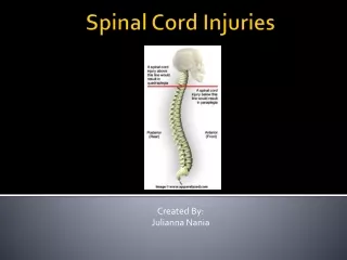
Anatomy • 33 vertebrae: 7 C, 12 Thoracic, 5 Lumbar, 5 sacral, 4 coccygeal • Denis three-column system for classification of thoracolumbar injuries • Anterior: ant vertebral body, ant. annulus fibrosus, ant long. Lig. • Middle: posterior wall of vert. body, post annulus fibrosus, post long. Lig • Posterior: post vertebral arch **if greater than two columns injured [unstable] **if greater than 50% compression [unstable]
Classification of spinal column injuries • Classified by mechanism • Flexion, flexion-rotation, extension, vertical compression • Flexion: Atlanto-occipital or atlantoaxial joint dislocation; simple wedge fracture; flexion teardrop fracture; clay shoveler’s fracture; bilateral facet dislocations
Damage to the corticospinaltract neurons (upper motor neurons) in the spinal cord results in (contralateral / ipsilateral) clinical findings such as muscle weakness, spasticity, increased deep tendon reflexes, and a Babinski sign.
When the _tract is damaged in the spinal cord, the patient experiences loss of pain and temperature sensation in the contralateral half of the body. • The (pain and temperature) sensory loss begins one or two segments below the level of the lesion.
Injury to one side of the dorsal • columns will result in (contralateral/ipsilateral) loss of vibration and position sense.
beginning with Tl, nerve roots exit (above/below) the vertebral body for which they are named.
Classification of spinal column injuries • Shear: odontoid fractures
Classification of spinal column injuries • Rotation: Rotary atlantoaxial dislocation; Unilateral facet dislocations
Classification of spinal column injuries • Extension: Posterior neural arch fracture of C1; hangman’s fracture; extension teardrop fracture
Classification of spinal column injuries • Vertical Compression: Compression fractures
Neurologic Evaluation • MOTOR EXAMINATION: • C4 Spontaneous breathing • C5 Shrugging • C6 Elbow Flexion • C7 Elbow Extension • C8-T1 Flexion of fingers • T1-T12 Intercostal & Abdominal muscles • L1-L2 Hip Flexion • L3 Hip Adduction • L4 Hip Abduction • L5 Foot Dorsiflexion • S1-S2 Foot Plantar flexion • S2-S4 Rectal Sphincter Tone
Neurologic Evaluation • Spinal Reflex Examination • C6 Biceps • C7 Triceps • L4 Patellar • S1 Achilles
Neurologic Evaluation • Spinal Sensory Exam • C2 Occiput L4 Knee • C3 Thyroid Cartliage L5 Lateral Aspect of Calf • C4 Suprasternal Notch S1 Lateral Aspect of Foot • C5 Below Clavicle S2-4 Perianal Region • C6 Thumb • C7 Index Finger • C8 Small Finger • T4 Nipple Line • T10 Umbilicus • L1 Femoral Pulse • L2-3 Medial Aspects of Thigh
Neurologic Evaluation • Complete lesions: total loss of motor & sensation • Spinal shock may mimic • May last several days; bulbocavernosus reflex marks end of shock • Sacral sparing • Perianal sensation, normal rectal sphincter tone, flexor toe movement
Neurologic Evaluation • Incomplete Spinal Lesions: • 90% are of three syndromes • Central Cord • Brown-Sequard • Anterior Cord
Neurologic Evaluation • Other 10% • Posteroinferior cerebellar artery syndrome – dysphageia, dysphonia, hiccups, vertigo, & cerebellar ataxia • Horner’s – cervical sympathetic chain damage with ipsilateral ptosis, miosis, and anhidrosis • Cauda equina – perineal or bilateral leg pain, bowel/bladder dysfunction, perianal anesthesia, diminished rectal tone, & lower extremity weakness • SCIWORA
Radiography • Nexus: prospective study 34,069 patients @ 21 EDs; all but 8 of 818 patients with injuries and only one required surgical stabilization • Criteria • No midline cervical tenderness • No focal neurologic deficit • No intoxication • Normal Alertness • No painful distracting injury
Radiography • Canadian Decision Rule: 3 questions • Are there any high-risk factors that mandate radiography? • Are there any low-risk factors that allow safe assessment of range of motion? • Is the patient able to actively rotate the neck 45 degrees to the left & right? • High Risks – age > 65 years; mechanism (fall > 1 m, an axial load, MVC > 100km/hr, rollover, ejection, ATV or bicycle collision); paresthesias • Low Risks – rear-end crashes, ability to sit up in ED, ability to ambulate, delayed onset of neck pain, absence of midline neck tenderness
Radiography • Cross-Table Lateral View • Three anatomical lines may be traced: • Along the anterior vertebral body cortex • Along the posterior vertebral body cortex • Along the spinolaminar junction • 25% children have pseudosubluxation C2/3 no more than 2 mm • retropharangeal soft tissues • C1-4 4-7mm • C5-7 16-22mm
Radiography • Trauma Series • AP/Lateral/swimmer’s/oblique/odontoid
Radiography • Flex/Ext Views: • Ant. Or Post. Subluxation > 2mm on one view and not on the neutral view = ligament injury • Only done if normal mental status & exam, but still pain • Controversial with CT/MRI available
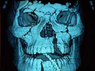
Radiography • MRI – superior for non-osseous eval. • CT • Indications – inadequate visualization; suspicious plain films; fracture/displacement on standard films; high clinical suspicion despite normal plain films • 3d reconstruction for complicated fractures
Unstable Fractures • Jefferson fracture • Hangman’s fracture • Flexion teardrop fracture • Extension teardrop fracture • Bilateral locked facets
Management • Assume injury • Immobilize • Watch out for spinal shock • Steroids? • Definitive Care