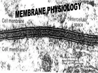
MEMBRANE PHYSIOLOGY
MEMBRANE PHYSIOLOGY. ARJUN MAITRA Asst. Professor Dept. of Physiology. LECTURE SLIDES FROM THE CLASSES TAKEN DURING 2008-09 SESSION. Topic: Cell membrane & Transport across the membrane Lecture taken for Courses: MBBS,BPT Useful for: B.ScNursing& others.

MEMBRANE PHYSIOLOGY
E N D
Presentation Transcript
MEMBRANE PHYSIOLOGY ARJUN MAITRA Asst. Professor Dept. of Physiology
LECTURE SLIDES FROM THE CLASSES TAKEN DURING 2008-09 SESSION Topic: Cell membrane & Transport across the membrane Lecture taken for Courses: MBBS,BPT Useful for: B.ScNursing& others
TO MY STUDENTS HERE I HAVE TRIED TO SIMPLIFY THE HUGE SUBJECT WITH ANIMATIONS, DIAGRAMS, FLOW CHARTS & RELEVENT MCQs. DIFFERENT TEXT BOOKS AND REFERENCE BOOKS HAVE BEEN USED FOR PREPARING THE CONTENTS. REMEMBERTHESE SLIDES ARE NOT THE SUBSTITUTE OF YOUR TEXT BOOKS ANIMATIONS AND DIAGRAMS ARE COLLECTED FROM DIFFERENT WEBSITE SOLELY FOR EDUCATION PURPOSE.
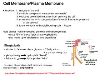
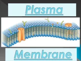
What is a Phospholipid? • It is a pair of fatty acid chains and a phosphate group attached to a glycerol backbone. • Polar (water-soluble) heads face out and the nonpolar fatty acids stay inside. IN OUT
cytoskeleton cholesterol carbohydrate chains membrane protein outside the cell interior of cell
Transport Proteins • Span the lipid bilayer • Interior is able to open to both sides • Change shape when they interact with • Solute • Play roles in active and passive transport
Carbohydrate layer (Glycocalyx) on the cell surface Protecting the cell surface from mechanical and chemical damage Lymphocyte stained with ruthenium red
Solutions • Solutions are made of solute and a solvent • Solvent - the liquid into which the solute is poured and dissolved. We will use water as our solvent today. • Solute - substance that is dissolved or put into the solvent. Salt and sucrose are solutes.
Weeee!!! high low This is gonna be hard work!! high low Types of Cellular Transport • Passive Transport cell doesn’t use energy • Diffusion • Facilitated Diffusion • Osmosis • Active Transport cell does use energy • Protein Pumps • Endocytosis • Exocytosis
Diffusion Diffusion: random movement of particles from an area of high concentration to an area of low concentration. (High to Low
Fick's law of diffusion J = - DA(DC/DX) where J is the net rate of diffusion, D is the diffusion coefficient, D = - ½b2f A is the area, and Δc/Δx is the concentration gradient. The minus sign indicates the direction of diffusion. TYPICAL VALUES OF THE DIFFUSION COEFFICIENT
Principles of Diffusion • Diffusion is the random mixing of particles that occurs in a solution as a result of the kinetic energy of the particles. • Diffusion rate across plasma membranes is influenced by several factors: – Steepness of the concentration gradient – Temperature – Size or mass of the diffusing substance – Surface area – Diffusion distance
FacilitatedDiffusion diffusion of specific particles through transport proteins found in the membrane
Conformational change of a carrier protein Mediates passive transport Change is spontaneous and random, so dependent on concentration
Permeability of plasma membrane General principles I
Permeability of plasma membrane General principles II Permeability coefficient (cm/sec)
Osmosis: diffusion of water through a selectively permeable membrane • Water moves from high to low concentrations
Active Transport • celluses energy • actively moves molecules to where they are needed • Movementfrom an area of low concentration to an area of high concentration • (Low High)
ACTIVE TRANSPORT PRIMARY ACTIVE TRANSPORT SECONDARY ACTIVE TRANSPORT
3 ways of driving active transportation utilizing passive carriers • Coupled carriers • ATP-driven pumps • Light-driven pumps
3 types of carrier-mediated transport Coupled carriers
Active transport enzymes couple net solute movement across a membrane to ATP hydrolysis. An active transport pump may be a uniporter or antiporter.
ATP-dependent ion pumps are grouped into classes based on transport mechanism, as well as genetic & structural homology. Examples include: • P-class pumps • F-class (e.g., F1Fo-ATPase to be discussed later) & related V-class pumps. ABC (ATP binding cassette) transporters, which catalyze transmembrane movements of various organic compounds including amphipathic lipids and drugs, will not be discussed here.
P-class ion pumpsare a gene family exhibiting sequence homology. They include: • Na+,K+-ATPase, in plasma membranes of most animal cells is an antiport pump. It catalyzes ATP-dependent transport of Na+ out of a cell in exchange for K+ entering. • (H+, K+)-ATPase, involved in acid secretion in the stomach is an antiport pump. It catalyzes transport of H+ out of the gastric parietal cell (toward the stomach lumen) in exchange for K+ entering the cell.
P-class pumps (cont): • Ca++-ATPases, in endoplasmic reticulum (ER) and plasma membranes, catalyze ATP-dependent transport of Ca++ away from the cytosol, into the ER lumen or out of the cell. Some evidence indicates that these pumps are antiporters, transporting protons in the opposite direction. Ca++-ATPase pumps function to keep cytosolic Ca++ low, allowing Ca++ to serve as a signal.
Na+-K+ Pump, ATPase P-type transport ATPase (dependent on phosphorylation)
Molecular weight Alpha Subunit 100,000 Beta Subunit 55,000 b • Alpha Sub unit • Span cell membrane for 10 times • NH2 and COOH both terminal • Intracellular • Phosphorylation site Asp 376 • Heterogenicity exists • Three isoforms are • a1 ----- membrane • a2--- muscle,heart,adipose tissue,brain • a3---- heart, brain a
Beta Subunits • Single membrane spanning unit • 3 extracellular glycolisation sites • Heterogenicity exists • 3 iso forms • b1---absent in astrocytes,vestibular cells, glycolytic fast twich muscles • b2 ---fast twich muscles • Beta 3 ----- not reported
EXOCYTOSIS • TWO PATHWAYS: • NONCONSTITUTIVE PATHWAY (SLOW) • CONSTITUTIVE PATHWAY (RAPID)
KISS AND RUN DISCHARGE • SNARE = SYNAPTOSOMAL ASSOCIATED RECEPTOR • FOR EXOCYTOSIS(soluble NSF attachment • receptor) • t-snare & v-snare NSF =N-ethylmaleimide-sensitive factor SNAP = soluble NSF attatchment protein