Download
1 / 22
950 likes | 4.24k Vues
Concentration methods of fecal parasites. Amal Almuhana 2012. Concentration techniques. A concentration technique is performed mainly to separate the parasites from fecal debris.

E N D
Concentration methods of fecal parasites AmalAlmuhana 2012
Concentration techniques • A concentration technique is performed mainly to separate the parasites from fecal debris. • The concentration procedure not only increases the number of parasites in the sediment but it also unmasks them, makingthem more visible by removing organic and inorganic debris.
Advantages &Disadvantages 1- Advantages • Maximizes the numbers of organisms detected which may be too scanty to be seen by direct microscopy alone . • separate the parasites from fecal debris. 2- Disadvantages • Destroys trophozoite stages and distorts cellular exudate .
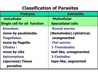
Types of Concentration techniques • There are two types of fecal Concentration techniques:- • Sedimentation techniques. • Flotation techniques.
1- Sedimentation techniques • Fecal sedimentation is used to detect large or heavy ova such as many spirurid ova, many fluke eggs, and many tapeworm eggs that will not float in fecal flotation techniques. • The sediment contains a lot of debris so examining these slides is quite tedious. Note:- you can use formalin as a preservative and ether or ethyl acetate as an extractor of fat and debris from faeces after filtration to leave the parasites in a sediment at the bottom of the tube after centrifugation.
Advantages &Disadvantages 1-Advantages • Recovers most ova, cysts and larvae . • Morphology of most parasites is retained. • Less risk of infection from bacteria and viruses. 2- Disadvantages • Preparation contains debris. • Some parasites do not concentrate well. • Ether is flammable and formalin is an irritant . • Liquid faeces do not concentrate well .
Materials • Stool samples • Glass slides • Coverslips • Pipettes • Stick • Gloves • Microscope • Plastic container (with small amount of water). • Centrifuge. • Centrifuge tube
Procedure 1- Obtain about a one to two gram fecal sample 2-Mix it with water by stick. 4-Pour the strained preparation into a centrifuge tube. 3- Strain the mixture
Procedure 5- Here is the appearance of the suspension before centrifugation. 6- Centrifuge at 1500 rpm for 5 minutes. 7-The appearance of the suspension after centrifugation. 8- Decant the supernatant
Procedure (cont.) 9- A few drops of the suspension will remain in the tube over the sediment pellet 10- Using a pipette remove some of the sediment from the top layer and place a drop on a slide for examination. 11-Place a cover slip over the drop of sediment suspension 12- Examine under microscope.
Procedure (cont.) • Note :-There are many variations on fecal sedimentation technique, many of which include a step using organic solvents (instead of water) to remove some of the material from the sediment. • Note:- at step (11):- • A drop of stain of your choice could be mixed in before placing the cover slip. • New Methylene blue or iodine solution enhances visualization of the parasite ova and oocytes. • If there is a lot of debris, water may be added first.
Examples of parasitic stages that might be seen under the microscope Entamoeba cysts. Faciolaegg
2- Flotation techniques • A fecal flotation test uses the high specific gravity of a solution to float the lighter ova and cysts • It is a diagnostic test commonly performed in-house in most veterinary clinics as a way of diagnosing parasitism in animals.
Advantages &Disadvantages 1- Advantages:- • The concentrate is clear of debris 2- Disadvantages:- • Delay in examination can result in distortion • Larvae and some fluke eggs do not concentrate • Frequent checking of specific gravity
Materials • Stool samples • Glass slide • Coverslips • fecal flotation test kit • Fecal floating solution. • Microscope • Gloves
Materials (cont. ) The FECALYZER (fecal flotation test kit). • The in-house fecal flotation 'Fecalyzer' (also spelled Fecalyser) apparatus. • used by many veterinarians, consists of an outer casing (the white plastic capsule or outer casing) containing a green fecal filtration 'basket'.
How to use the FECALYZER?(procedure ) 1- Remove the inner piece of the fecal float test kit and press it into the fecal sample until the end is full. 2- Place insert back inside the test container. 3- Locate the fill line on the side
Procedure cont. 4- Fill insert with fecal solution until it gets to the top. 5- Carefully add just enough solution to form a meniscus on top without allowing it to over flow. 6-Gently place the slide cover slip on top of the insert and let it sit for approximately 12 to 15 minutes.
Procedure cont. 7-The glass coverslip (containing fecal float solution and, parasite (eggs/oocystes) is placed on top of the glass microscope slide. 8- Examine slide under microscope
Examples of parasitic stagesthat might be seen under the microscope Isosporacanis (coccidia) oocysts seen at low power Bacterial rods seen at 400x. Isosporaoocyst(extreme close-up - 1000x) . Coccidia are single-celled organisms
The important points to be considered when performing a concentration technique are: • In a specimen the whole of the sample (equivalent to 1 gram of faeces) should be concentrated and the whole of the deposit examined. • It is important to vortex the sample for at least 15 seconds after the addition of ether or ethyl acetate thus obscuring ova and cysts. • Adequate centrifugal force must be used because if this is below the required value, there may be insufficient gravitational force to sediment the ova and cysts. • The centrifugal time is also critical, since the ova and cysts may remain in suspension if the sample is not centrifuged for the minimum required time.