Yoke muscles
Yoke muscles. Agonist, antagonist and yoke muscles. Agonist. Agonist, antagonist and yoke muscles. Agonist Muscle that moves the eye in a particular direction Example?. Agonist, antagonist and yoke muscles. Agonist

Yoke muscles
E N D
Presentation Transcript
Agonist, antagonist and yoke muscles • Agonist
Agonist, antagonist and yoke muscles • Agonist • Muscle that moves the eye in a particular direction Example?
Agonist, antagonist and yoke muscles • Agonist • Muscle that moves the eye in a particular direction For example, the lateral rectus is the agonist for abduction or lateral gaze
Agonist, antagonist and yoke muscles • Agonist • Antagonist
Agonist, antagonist and yoke muscles • Agonist • Antagonist • Muscle causing movement in direction opposite that of agonist Example?
Agonist, antagonist and yoke muscles • Agonist • Antagonist • Muscle causing movement in direction opposite that of agonist For example, the lateral and medial recti are antagonists for lateral gaze
Agonist, antagonist and yoke muscles • Agonist • Antagonist • Synergist
Agonist, antagonist and yoke muscles • Agonist • Antagonist • Synergist Two muscles moving the eye in the same direction. Example?
Agonist, antagonist and yoke muscles • Agonist • Antagonist • Synergists Two muscles moving the eye in the same direction. For example, the superior oblique and the inferior rectus are synergistic for downward gaze. These muscles are NOT synergists for every action. For torsion, they are antagonists.
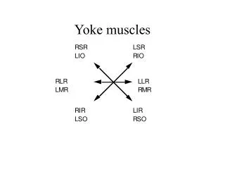
Agonist, antagonist and yoke muscles • Agonist • Antagonist • Synergist • Yoke muscles
Agonist, antagonist and yoke muscles • Agonist • Antagonist • Synergist • Yoke muscles are the synergist muscles in the two eyes, those that cause the two eyes to move in the same direction.
Agonist, antagonist and yoke muscles • Agonist • Antagonist • Synergist • Yoke muscles are the synergist muscles in the two eyes, those that cause the two eyes to move in the same direction. For example, RSO and LIR are yoke muscles in depression in left gaze
Hering’s law of equal innervation • Corresponding (or yoke) muscles of the two eyes must receive equal innervation (or input) so two eyes move together and by the same amount (Hering, 1868) • The yoke is neural, not mechanical.
Will the eyes always move together? • Not if there is a paralysis of muscle(s) in one eye • Here, equal inputs to the two eyes will not have equal effects because one or more muscles is weak • Illustration: case of recent paralysis of right lateral rectus
Recent paralysis of RLR • When the left eye fixates, both eyes should receive normal input • This will not result in normal contraction of the palsied muscle • The right eye should look to the left of the point of fixation • This is the primarydeviation of the palsied eye.
Recent paralysis of RLR • When the right eye fixates, brain must send supernormal input to RLR and simultaneously, supernormal input to its yoke muscle, LMR. • Now the left eye will turn too far to the right. • This is called secondary deviation • greater than the primary deviation due to Hering’s Law.
Law of reciprocal innervation • Sherrington: Increases in contraction do not proceed simultaneously in antagonist muscles. • Instead, as one muscle of an antagonist pair contracts, the other relaxes.
Proof of reciprocal innervation in extraocular muscle • Cut Cranial Nerves III and IV on the right side • in the right eye, only the lateral rectus muscle had normal input and normal tonus • Because RLR action was unopposed by any input to its antagonist, the right eye was abducted when at rest.
Proof continued • With Cranial Nerves III and Ivcut, stimulate cerebral cortex to produce levoversion (conjugate left eye movements • Predicted result: increased input to the left LR and to the right MR • Because the IIIrd nerve had been cut, the MR muscle does not receive this input.
End of proof • Principle of reciprocal innervation predicts: during levoversion, there is decreased input to LMR and RLR • OD will move left (to the midline) because the LR relaxes
Other evidence for reciprocal innervation • Modern studies with the electromyogram (EMG) • As activity in the antagonist decreases, the agonist increases
Combined Actions of the Muscles • During any movement of the eyes all of the muscles participate in some manner. • Some muscles move the eye (agonists) and so contract while others, those opposing the movement (antagonists) are relaxed. • Examples from a handout by Dr. Christensen
Abduction • LR (agonist) receives input from brain and contracts starting to move the eye outward. • MR (antagonist) is inhibited and so relaxes. • SO and IO (synergists for abduction) will contract to negate each others torsional effects. • SR and IR (weak anatonists) will relax and will also negate each others torsional effects.
Abduction • With the eye near primary position, the abductive function of the superior and inferior obliques is opposed by the adductive potentialities of the superior and inferior rectus. • Once the eye has been abducted more than 23°, both the vertical recti and the obliques assist in the abduction. • With extreme abduction, the lateral rectus is less important as an abductor as medial rectus tension increases. The assistance of the obliques and the vertical recti is crucial.
Elevation and Abduction • LR, SR and the IO contract while MR, IR and the SO relax. • LR and IO abduct with SR countering initially; later it aids in the abduction. • From primary position SR and IO initiate elevation. • In extreme abduction, only the SR elevates. • IR and SO relax equally to counter unwanted torsions.