Mesenteric adenitis in children
Mesenteric adenitis in children. Geetha M Pediatric Gastroenterologist Amrita Hospital, Cochin. Scenario. Mesenteric Lymphadenopathy not a diagnosis Incidental finding in Recurrent Abdominal Pain USG abdomen is one primary investigation Organic causes 4-11%

Mesenteric adenitis in children
E N D
Presentation Transcript
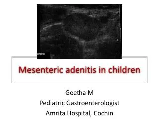
Mesenteric adenitis in children Geetha M Pediatric Gastroenterologist Amrita Hospital, Cochin
Scenario • Mesenteric Lymphadenopathy not a diagnosis • Incidental finding in Recurrent Abdominal Pain • USG abdomen is one primary investigation • Organic causes 4-11% • USG findings- Mesenteric nodes, GB Stones • ? Significance
MLN • Medical literature • Pediatric Literature – specific inflammation by Yersinia, Staph, Salmonella • Radiological literature - LN > 5mm size • What is the significance? Importance of Sonographic Detection of Enlarged Abdominal Lymph Nodes in Children Natalia Simanovsky, MD, Nurith Hiller, MD J Ultrasound Med 2007; 26:581–584
What is mesenteric adenitis? • 3 or > LN • 4 mm or > in short axis: 8mm > in long axis • Primary- when LN are the only finding • Secondary – when another pathology is identified • Incidence varies Rao PM, Rhea JT, Novelline RA. CT diagnosis of mesenteric adenitis. Radiology 1997; 202:145–149.
Causes - Local Infections • Gastroenteritis • Appendicitis • Parasitic infections • IBD
Parasitic Infection • Parasitic infec is a cause of RAP • ?? Cause for MLN?? • 2002-2008 , 224 children with RAP • 89 boys: 135 girls ; Mean age 9 yrs • Pedsonologist • Short axis >8mm = enlarged MLN Enlarged mesenteric lymph nodes in children with recurrent abdominal pain: Is there an association with intestinal parasitic infections? FraukjeWiersma et al
Contd…….. • All children had MLN at least 5mm • 86% (193/224) - had all nodes < 5mm • 6/224 (2.5%) > 8mm: 25/224 (11.2%) 5-7mm • None of the 6 had parasites • 25% (56) had parasitic infection • 47 - < 5mm • 9 – 5-7 mm • Concluded – not related to parasitic infection Simanovsky N et al. Importance of sonographic detection of enlarged abdominal lymph nodes in children. J Ultrasound Med 2007; 26:581-584
Infections associated with MLN • Yersiniaenterocolitica - RIF syndrome • Atypical Mycobacteria • Campylobacter spp • Coxackie virus, EBV • HIV Jelloul I, Fremond B, Dyon JF, Orme RI, Babut JM. Mesenteric adenitis caused by Yersiniapseudotuberculosis presenting as abdominal mass. Eur J Pediatr Surg 1997; 7:180–183. Nilehn B, Sjostrom B. Studies on Yersiniaenterocolitica. Occurrence in various groups of acute abdominal disease. ActaPatholMicrobiol Scand 1967; 71:612-628.
Symptomatology • Mostly asymptomatic • Diffuse abd pain – sometimes localised in RLQ • Concomittant/ antecedent URI • Anorexia • Diarrhoea • Nausea/ vomiting
Symptoms………….Contd • Fever • Rhinorrhoea • RLQ tenderness • 20% peripheral lymphadenopathy • LN Biopsy – mostly reactive/ non – specific inflammation
Early Studies • LN > 4mm in AP diameter – 4% asymp children • 10-20 mm long axis 89% asymp children • MLN (long axis) in almost all children Sivit CJ, et al. Visualization of enlarged mesenteric lymph nodes at US examination. PediatrRadiol 1993; 23:471-475 Healy MV, Graham PM. Assessment of abdominal lymph nodes in a normal pediatric population: an ultrasound study. AustralasRadiol 1993; 37:171–172. Watanabe M, Ishii E, Hirowatari Y, et al. Evaluation of abdominal lymphadenopathy in children by ultrasonography. PediatrRadiol 1997; 27:860–864
CT and MLN • All non contrast CT images done for renal stones were evaluated for MLN • 33/61 had MLN mostly in RLQ • Max size 10 mm – also in RLQ • Cluster of 3 nodes – RLQ • 5mm size nodes – in almost all • Hence a measurement of 8mm or > chosen Karmazyn B, Werner EA, Rejaie B, Applegate KE. Mesenteric lymph nodes in children: what is normal? PediatrRadiol 2005; 35:774-777
Which size is significant ? • MLN in children – asymptomatic and RAP • 200 children • Acute abd / RAP/ others • Only > 10 mm was statistically significant Importance of Sonographic Detection of Enlarged Abdominal Lymph Nodes in Children Natalia Simanovsky, MD, Nurith Hiller, MD. J Ultrasound Med 2007; 26:581–584
Does Size Matter ? • LN > 4mm seen in 4-64% asymp children • 14-83% of symp children • MLN are seen in all children – asymp, symp- acute abd, CAP, gastroenteritis • Tendency to have larger nodes in acute infect • As an isolated finding – not much importance Sivit CJ, et al. Visualization of enlarged mesenteric lymph nodes at US examination. PediatrRadiol 1993; 23:471-475 Rathaus Vet al Enlarged mesenteric lymph nodes in asymptomatic children: the value of the finding in various imaging modalities. Br J Radiol 2005; 78:30-33 Nan Fang Yi KeDaXueXueBao. 2011 Mar;31(3):522-4. [Enlarged mesenteric lymph nodes in children: a clinical analysis with ultrasonography and the implications]. [WANG WG, TIAN H, YAN JY, LI T, ZHANG TD, ZHAO YP, ZHANG LY, XING HG.
Distribution of EALNs of 5 mm or larger in the shortest diameter by age Importance of Sonographic Detection of Enlarged Abdominal Lymph Nodes in Children Natalia Simanovsky, MD, Nurith Hiller, MD. J Ultrasound Med 2007; 26:581–584
Indian Experience • MLN almost universally seen • Enlarged nodes > 8mm upto 20mm • If isolated and clinically well – only follow up • If symptomatic - course of antibiotics • Usually pain tends to settle but nodes persist • If persistent and symptomatic - evaluate
Conclusions • Frequent in asymptomatic children • Nodes 10 mm or > in setting of abdominal pain – considered as ML • Usually increase in size till 10 yrs and then regress • Mostly non specific – but follow up if necessary