Download

1 / 27
280 likes | 485 Vues
The Spine and Its Muscles. Terms . Capitis = head Cervicis = neck Thoracis = thoracic Lumborum = Lumbar Foramen = hole/tunnel Occiptal condyles = where C1 articulates. Thorax/Ribs. Thorax = refers to entire chest

E N D
Terms • Capitis = head • Cervicis = neck • Thoracis = thoracic • Lumborum = Lumbar • Foramen = hole/tunnel • Occiptal condyles = where C1 articulates
Thorax/Ribs Thorax = refers to entire chest 12 pairs of ribs give structural support to the thoracic cavity 1-7 have direct attachment to the sternum by costalcartilage, making them true ribs. The other 5 ribs are false ribs because they either attach indirectly or not at all to the sternum. 11-12 ribs are floating because they do not attach to the sternum at all
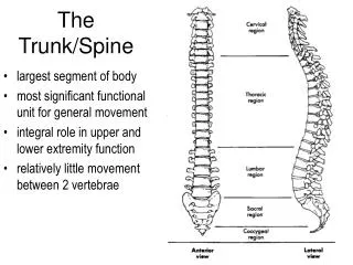
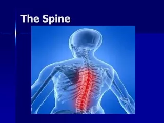
Functions of Spine • The vertebral column is a strong, flexible rod that bends anteriorly, posteriorly, laterally, and rotates. • The spine serves to protect the spinal cord, support the head, and is a point of attachment for ribs, pelvic girdle and muscles of the back. • The spine makes up about 2/5 of the total height of the body.
Vertebrae • Between vertebrae are openings called intervertebral foramen. The spinal nerves pass through these openings, and connect to various parts of the body. • The adult vertebrae is divided into 5 regions that contain 33 bones: • Cervical = 7 vertebrae *Think of your own way • Thoracic = 12 vertebrae remember these: • Lumbar = 5 vertebrae ____________________ • Sacrum = 5 fused vertebrae • Coccyx = 4 fused vertebrae
Intervertebral Discs • Discs lie in between the bodies of adjacent vertebrae. • The outer fibrous ring is call the annulus fibrosis. • The inner part of the disc is filled with “jelly” substance, and is the nucleus pulposus. • The discs serve to form joints, permit movement, and absorb shock. * Can a disc “slip?” What happens during this?
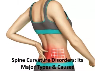
Curves of the spine • The cervical and lumbar curves are anteriorly convex= lordosis (give an example: ) • The thoracic and sacral curves are anteriorly concave = kyphosis (give an exammple: ) • The curves are important because they increase strength, help maintain balance, absorb shock, and help protect the spinal column from fracture. • In the fetus there is only a single kyphosis curve, but approximately the third month after birth the cervical curve develops. • When the child sits up, stands, and walks, the lumbar curve develops. • All curves are fully developed by age 10.
Parts of Vertebrae • Vertebrae consist of a body, vertebral arch, and several processes. • The body is the weight bearing part of a vertebrae. • The arch is formed by the pedicles and the laminae, which serve to create the vertebral foramen. • The vertebral foramen protects the spinal cord. • When the vertebrae arestackedon each other, it creates an opening between adjoining vertebrae on each side of the spinal column. • These openings are the intervertebral foramen, and allow for each spinal nerve to pass through. • The cervical nerve roots exit above the vertebrae, whereas the thoracic and lumbar exit below. • Why is there a C8 nerve, if we only have 7 cervical vertebrae? • Spinal cord ends at about L2, then gives rise to the caudaequina(horse tail)
Vertebrae continued • Each vertebrae have a spinousprocess, which can be felt on the backof your spine. • The spinous process serves as an attachment point for muscles. • The facets are articulating surfaces that form the joints of the spine. They allow for movement of the spine.
Cervical Vertebrae • Atlas & Axis = first two cervical vertebrae • The Atlas articulates with the skull is the “yes” joint • The Axis sits below the atlas the “no” joint • Have horizontal facetsthat allow for rotation of the head • Have a transverse foramen for arteries to pass through • Have a bifid (forked) spinous process
Thoracic Vertebrae • Have longerspinous process • Have multiple facets to articulate with other vertebrae above and below, and also with ribs • Have posterior facing facets that allow for rotation
Lumbar Vertebrae • The largest and strongest in the vertebral column because of the amount of body weight they have to support • The spinous processes have attachments of large back muscles • Have medial/lateral facing facets to allow for flexion of the spine
Ligaments • Anterior longitudinal Ligament = anterior aspect of vertebral bodies stops extension of spine • Posterior longitudinal ligament = runs along the posterior aspect of vertebral bodies stops flexion of spine ….. Is more narrow than the anterior ligament, which causes less stability during flexion of the spine (this is the reason why most disc bulges are posterior) • Ligamentumflavum = between lamina
Muscles • Transversospinalis group (semispinalis) = extension and rotation of spine • Multifidus= extension and rotation of spine • Rotatores= extends and rotates spine • Intertransversarii = stabilzer • Interspinalis = stabilizer
Sternocleidomastoid (SCM) • O: Manubrium of sternum = medial 1/3 of clavicle • I: Mastoid Process of temporal bone • Innervation: Cranial Nerve XI – Spinal Accessory Nerve
Sternocleidomastoid (SCM) • Action • Together: Flex head and neck • Separate: Lateral Flexion ipsilaterally + Rotate, extend contralaterally • Torticollis
Primary Movers Trunk
Interal + External Obliques • Internal • O: Middle lip iliac crest • I: Inferior border lower ribs, fuses with external oblique to form rectus sheath • External • O: Outer surface lower ribs • I: Outer iliac crest, rectus sheath • Innervation: thoracic + lumbar nerves • Action: Flexes trunk, lateral flexion ipsilaterally, Erotates contralaterally, Irotates ipsilaterally
Rectus Abdominis • O: Costal cartilage ribs 5-7, xiphoid process • I: Pubis • Innervation: Lower thoracic nerves • Flexes trunk, compresses abdominal wall
Erector Spinae Group • Iliocostalis • Lumborum • Thoracis • Cervicis • Longissimus • Thoracis • Cervicis • Capitis • Spinalis • Thoracis • Cervicis • Capitis • Action: • Primary back + head extensors • Rotate head ipsilaterally • Primary lateral flexors of spine
Primary Stabilizers Muscles that help prevent back injury – THE CORE!
Transverse Abdominis • O: Inner lip iliac crest, inner surface lower ribs • I: Linea Alba, pubic rim, rectus sheath • Innervation: Lower thoracic and upperlubar nerves • Action: Tenses abdominal wall, stabilizes lumbar spine
Transversospinalis: Multifidus • From lateral to transverse process above • Span 2-4 vertebrae • Best developed in lumbar region • Action
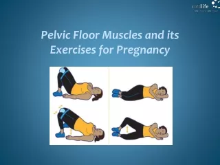
Transversus abdominis, multifidis, pelvic floor muscles • Segmental stabilizers of low back – “natural weight belt” • Pre-movement activated, shut down with injury • SAID training only
Core Muscles Most superficial to most deep: • Rectus abdominis(six pack muscle) • External oblique • Internal oblique • Transverse abdominis= works with the multifidus to support the spine all the way around