Download
1 / 54
540 likes | 1.04k Vues
6. Obtaining Vital Signs and Measurements. Learning Outcomes. 6.1 Describe vital signs and common body measurements. 6.2 Differentiate measurement systems. 6.3 Identify the instruments used to measure vital signs and body measurements.

E N D
6 Obtaining Vital Signs and Measurements
Learning Outcomes 6.1 Describe vital signs and common body measurements. 6.2 Differentiate measurement systems. 6.3 Identify the instruments used to measure vital signs and body measurements. 6.4 Carry out vital signs and body measurements of infants, children, and adults.
Learning Outcomes (cont.) 6.5 Recognize abnormal vital signs and body measurements. 6.6 Write vital signs and body measurements using accurate terminology and abbreviations. 6.7 Implement growth charts.
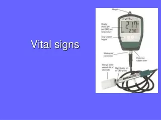
Vital signs Temperature Pulse Respirations Blood pressure Pain assessment Body measurements Height Weight Head circumference Introduction Vital signs and body measurements are used to evaluate health problems. Accuracy is essential.
Vital Signs • Provide information about patient’s overall condition • Taken at each visit and compared to baseline • Protected health information – HIPAA
Temperature • Febrile – body temperature above patient’s normal range • Fever – sign of inflammation or infection • Hyperpyrexia – extremely high temperature • Afebrile – normal body temperature
Ear Mouth Rectal Axilla Temporal Artery Temperature (cont.) • Measurements • DegreesFahrenheit(°F) • Degrees Celsius(centigrade;°C) • Normal adult oral temperature • 98.6°F • 37°C Temperature Routes
Thermometers • Electronic digital thermometer • Accurate, fast, easy to read • Comfortable for the patient • Tympanic thermometer • Temporal scanner • Disposable thermometer • Single use • Less accurate Disposable sheaths are used with electronic thermometersto prevent cross-contamination.
Taking Temperatures • Measure to nearest tenth of a degree • Oral temperatures • Wait at least 15 minutes after eating, drinking, or smoking • Place under tongue in either pocket just off-center in lower jaw
Taking Temperatures (cont.) • Tympanic temperatures • Proper technique essential • Adult – pull ear up and back • Child – pull ear down and back • Fast, easy to use, and preferred in pediatric offices
Taking Temperatures (cont.) • Rectal temperatures • Use Standard Precautions – gloves • Patient is positioned on side (left side preferred) or stomach • Lubricate tip of thermometer • Slowly and gently insert tip into anus • ½ inch for infants • 1 inch for adults • Hold thermometer in place while temperature is taken
Axillary temperatures Place patient in seated or lying position Place tip of thermometer in middle of axilla with shaft facing forward Probe must touch skin on all sides Temporal temperatures Temporal scanner Noninvasive, quick Stroke scanner across forehead, crossing over the temporal artery Taking Temperatures (cont.)
Taking Temperatures (cont.) • Children • Take temperature last if child cries or becomes agitated • Agitation will cause pulse, respiration, and blood pressure to elevate • Oral route is not appropriate for children under 5 years old
Respiratory Respirations Circulatory Pulse Pulse and Respiration Linkage Pulse and respirations are related because the heart and lungs work together. Normally, an increase or decrease in one causes the same effect on the other.
Pulse • Pulse – number of times the heart beats in 1 minute • Respiration – number of times a patient breathes in 1 minute • One breath = one inhalation and one exhalation • Ratio of pulse to respirations is 4:1
Indirect measurement of cardiac output Problems if Tachycardia Bradycardia Weak Irregular Sites of measurement Adults – radial artery Children – brachial artery(antecubital space) Apex of heart 5th intercostal space directly below center of left clavicle Apical pulse taken with a stethoscope Pulse (cont.)
Pulse (cont.) • Locate pulse by pressing lightly with index and middle finger pads at the pulse site • Count the number of beats felt in 1 minute • If regular – may count beats for 30 seconds and multiply by 2
Pulse (cont.) Regular Pulse Rhythm Irregular Pulse Rhythm • Count for 30 seconds, then multiply by 2 (a rate of 35 beats in 30 seconds equals a pulse rate of 70 beats/minute) • Count for one full minute • May use stethoscope to listen for apical pulse and count for a full minute Click for sounds
Pulse (cont.) • Electronic measurement devices • Blood pressure machine • Pulse oximetry unit • Infrared light measures pulse and oxygen levels • Report oxygen level below 92% not improved by deep breathing
Respiration • Respiratory rate – indication of how well the body provides oxygen to the tissues • Check by watching, listening, or feeling movement 1 inhalation + 1 exhalation = 1 respiration
(26-40) (20-30) (18-24) (12-24) (16-24) (12-20) Normal Respiratory Rates NOTE: Ranges reflect breaths per minute
Check respirations Look, listen, and feel for movement of air Count with a stethoscope Count for one full minute Rate Rhythm – regular Effort (quality) – normal, shallow, or deep Respiration (cont.) NOTE: If patients are aware that you are counting respirations, they may unintentionally alter their breathing.
Respiration Irregularities (cont.) • Indication of possible disease • Hyperventilation – excessive rate and depth • Dyspnea – difficult or painful breathing • Tachypnea– rapid breathing • Hyperpnea – abnormally rapid or deep breathing
Respiration Irregularities (cont.) • Rales (noisy) • Constriction or blockage of bronchial passages • Pneumonia, bronchitis, asthma, or other pulmonary disease • Cheyne-Stokes respirations • Periods of increasing and decreasing depth of respiration between periods of apnea • Strokes, head injuries, brain tumors, congestive heart failure • Apnea – absence of breathing
Blood Pressure • The force at which blood is pumped against the walls of the arteries (mmHg) • Two pressure measurements • Systolic pressure – measure of pressure when left ventricle contracts • Diastolic pressure • Measure of pressure when heart relaxes • Minimum pressure exerted against the artery walls at all times
Systolic Pressure Diastolic Pressure • Contraction of left ventricle • Top or first number • Heart at rest • Bottom or second number Blood Pressure (cont.) 120/80
Factors Affecting Blood Pressure • Internal • Cardiac output • Blood volume • Vasoconstriction • Viscosity
Hypertension Benign – no risks to other organs Malignant – with other conditions such as renal or heart failure Hypotension Not generally a chronic health problem Severe hypotension may present with shock, heart failure, severe burns, excessive bleeding Factors Affecting Blood Pressure (cont.)
Sphygmomanometer Inflatable cuff Pressure bulb or other device for inflating cuff Manometer Types Aneroid Electronic Mercury BP Measurement Equipment (cont.)
Measurement Equipment (cont.) • Aneroid sphygmomanometers • Circular gauge for registering pressure • Each line 2 mmHg • Very accurate • Must be checked, serviced, and calibrated every 3 to 6 months
Measurement Equipment (cont.) • Electronic sphygmomanometers • Provides a digital readout of the blood pressure • No stethoscope is needed • Easy to use • Maintain equipment according to manufacturer’s instructions
Measurement Equipment (cont.) • Mercury sphygmomanometers • A column of mercury rises with an increased pressure as the cuff is inflated • No longer available for purchase • If in use, must be checked, serviced, and calibrated every 6 to 12 months
Measurement Equipment (cont.) • Stethoscope – amplifies body sounds • Earpieces • Binaurals and tubing • Chestpiece • Bell – low-pitched sounds • Diaphragm – high-pitched sounds Earpieces Binaurals Rubber or plastic tubing Bell Chestpiece Diaphragm
Place cuff on the upper arm above the brachial pulse site Inflate cuff about 30 mmHg above palpatory result or approximately 180 mmHg to 200 mmHg Slowly release the air in cuff and simultaneously listen for vascular sounds Korotkoff sounds – five phases Measuring Blood Pressure
Measuring Blood Pressure (cont.) • Korotkoff sounds • Phase 1 – tapping sound represents the systolic pressure • Phase 2 – softer swishing sound • Phase 3 – resumption of a crisp tapping sound • Phase 4 – sound changes to muffled • Phase 5 – sound disappears; represents the diastolic pressure • Record results with systolic as the top number and diastolic as the bottom number (i.e., 120/76)
Blood Pressure (cont.) • Special considerations in adults • Post exercise, ambulatory disabilities, obese, known blood pressure problems • Anxiety or stress • Avoid measurement in an arm • Injury or blocked artery is present • History of mastectomy on that side • Implanted device is under the skin • Proper cuff size – improper size results in inaccurate reading
Blood Pressure (cont.) • Special considerations in children • Not routinely taken on each visit • Take before other tests or procedures • Cuff size important • Palpatory method not used with children • Heartbeat may be heard to zero; record diastolic when strong heartbeat becomes muffled
Orthostatic or Postural Hypotension • Blood pressure becomes low and pulse increases when the patient moves from lying to standing • May indicate dehydration, heart disease, diabetes, medications, or nervous system disorder • Vital signs are taken in different positions • Positive tilt test– increase in pulse > 10 bpm and a drop in BP > 20 mmHg
Correct! Apply Your Knowledge • You are about to take the temperature of a 6-month-old infant being seen at the pediatrician’s office for vomiting and diarrhea. Which route will you use and why? What special considerations do you need to keep in mind with this specific patient situation and why? Answer: Route would be either tympanic or temporal since a 6-month-old would not be able to hold the thermometer under his/her tongue. Special considerations include: Taking the temperature after the pulse and respirations. For the tympanic thermometer, use proper technique and pull the ear down and back. Use Standard Precautions to prevent the spread of microorganisms.
Correct! Apply Your Knowledge • A 26-year-old athlete visits the medical office for a routine checkup. The medical assistant takes T-P-R and obtains the following: Temperature 98.8°F, Pulse 52 beats/minute, andRespirations 18/minute. What should the medical assistant do about these results? ANSWER: The temperature and pulse are within the normal range. The pulse of 52 is below the normal range. Check the patient’s previous vital sign results. Some patients, especially athletes, normally have a low pulse rate, so these results may be within normal limits for this patient.
Apply Your Knowledge • A 67-year-old patient is in the medical office complaining of a headache. The blood pressure reading is 212/142. What should the medical assistant do in this situation? ANSWER: This blood pressure reading is very high and should be reported to the physician at once. The complaint of headache should also be reported to the physician. Hypertension is a major contributor to stroke and heart attacks. 3 FOR 3! Very Good!
Adults and older children Height Weight Infant measurements Length Weight Head circumference Body Measurements Provide baseline values for current condition and enable monitoring of growth and development of children.
Adult weight Taken at each office visit Record to nearest quarter of a pound Height of adults Taken on initial visit and yearly thereafter Height bar on scale Record to nearest quarter of an inch Body Measurements (cont.)
Body Measurements (cont.) • Weight of children and infants • Children • Adult scales if able to stand • Held by an adult using the adult scale, and subtract adult weight from total to yield child’s weight • Infants • Infant scales
Body Measurements (cont.) • Height of children and infants • Children • Height bar on scale • Wall charts • Infants • Length measured at each visit • Built-in bar on exam table • Tape measure or yardstick
Body Measurements (cont.) • Head circumference of infants • An important measure of growth and development • Tape measure is placed around head at its largest circumference to obtain measurement
Body Measurements (cont.) • Other measurements • Diameter of limb – measure both to determine difference in size • Wound, bruise, or other injury – length and width to evaluate healing process • Chest circumference in infants • Abdominal girth in adults
Apply Your Knowledge The medical assistant is about to weigh a 6-month-old infant using the infant scale. When the medical assistant places the infant on the scale she notices the diaper is very soiled. What should the medical assistant do? ANSWER: The diaper could be changed prior to weighing. However, if the infant is weighed with the soiled diaper, the medical assistant should weigh the diaper after weighing the infant and subtract the difference to obtain the infant’s accurate weight. Correct!
6.1 Vital signs include temperature, pulse, respirations, blood pressure and assessment of pain. The most common body measurements are height, weight, and head circumference. 6.2 Mathematical formulas used to convert between Celsius and Fahrenheit and kilograms and pounds are: °F = ( °C X 9/5 + 32) [set fraction 9/5 on top] °C = ( °F – 32) X 5/9 [set fraction 5/9 on top] lbs = kb X 2.205 kg = lbs X 0.454 In Summary
In Summary (cont.) 6.3 Instruments used to measure vital signs and body measurements include a thermometer, temporal scanner, stethoscope, sphygmomanometer, scale, and tape measure. 6.4 The procedure to measure vital signs and body measurements is done with extreme care to ensure accuracy. Standard Precautions and aseptic technique must be utilized to prevent the spread of infection. Document information according to your facility policy.