Understanding Meiosis: The Process of Gamete Formation in Cellular Division
350 likes | 500 Vues
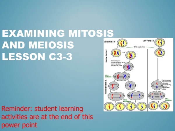
This lesson focuses on meiosis, the specialized form of cell division responsible for creating gametes (sperm and eggs). It covers the two stages of meiosis, Meiosis I and Meiosis II, each comprising distinct processes such as Prophase, Metaphase, Anaphase, and Telophase. The lesson also explains the significance of interphase and features a hands-on activity utilizing yarn to visualize chromosome duplication and movement. By the end, students will understand how diploid cells (2n) divide to form four haploid cells (n) essential for reproduction.

Understanding Meiosis: The Process of Gamete Formation in Cellular Division
E N D
Presentation Transcript
Unit: GeneticsLesson: Meiosis Agriscience Curriculum
Meiosis • Form of cell division that ultimately creates gametes – there are two types • 1. Sperm formation (called spermatogenesis) • 2. Ovum or Egg formation (called oogenesis)
Meiosis – the overview • In meiosis, there are two separate divisions, Meiosis I and Meiosis II. • By the end of both processes, the original diploid cell (2n) cell has divided into four haploid cells (n). • These haploid cells are called gametes: sperm and eggs. When fertilized, the resulting cell is called a zygote, again with a diploid number of cells.
Interphase – a step ahead of meiosis! • Prior to beginning meiosis I, the cell replicates its chromosomes in interphase. After replication, each chromosome consists of two identical sister chromatids, held together by a centromere. • Directions for story board: Take out a piece of yarn (each student). This is your original chromosome. Take out another piece of yarn identical to the first. This is your duplicated chromosome. Tie them together with twisty-tie. This twisty-tie is called the centromere. Glue them to the first block on the cardboard and label it “Interphase.” Your chromosome has just been duplicated!
All Chromosomes Replicate in Interphase! Prior to meiosis, all chromosomes are duplicated in a process similar to chromosome duplication prior to mitosis.
Next is Meiosis I ! Meiosis I contains four steps. • Prophase I • Metaphase I • Anaphase I • Telophase I
Prophase I 1. In Prophase I: a) Chromosomes appear, coil up and a spindle forms. b) Each chromosome pair comes together with another, and it matched up gene to gene. This creates a four-part structure called a tetrad. c) Nuclear envelope disappears. d) Crossing over occurs at site called chiasma. This is simply where genetic information is exchanged because the chromosome pairs are held so tightly together. This results in new allele combinations.
Directions for Storyboard • Have two students pair their duplicated chromosomes (done in interphase). • Connect these two chromosome pairs with another twisty-tie. • Glue these pairs to the second storyboard block and label the diagram as seen in slide #9. This step is called…. Prophase I
Metaphase I 2) Metaphase I: Pairs of homologous chromosomes move to mid-line of the cell as the centromeres (holding the chromosomes together) attach to the spindle fiber of the cell.
Directions for Storyboard • In block three of the storyboard, draw a circle to represent the cell membrane. • Through the center of the circle, glue a pipe-cleaner – this represents the spindle of the cell. • Take pairs of chromosomes and line them up across the spindle and glue them in. The centromeres (or twisty-ties) should be on the spindle (the pipe cleaners). • At the left and right borders of the circle, glue a button. These represent the centrioles, or poles of the cell.
Anaphase I 3) Anaphase I: a) One chromosome from each homologous pair of chromosomes move to opposite poles of the cell.
Directions for Storyboard • Separate the homologous pairs of chromosomes and have them “pulled” towards opposite centrioles (poles) of the cell. • Glue into your storyboard in box #4 and label it, “Anaphase I.”
Telophase I • Telophase I varies from species to species. Sometimes Telophase I is skipped and meiosis starts its second division immediately. In general, however, two nuclear envelopes begin to surround the separate chromosomes and cytokinesis (splitting of the cytoplasm into two separate entities) will sometimes occur. Then a phase called interkinesis will follow, which essentially is a resting period from Telophase I to Prophase II. This differs from mitosis because DNA replication does not occur.
Directions for Storyboard • Draw one circle in box 5 of your storyboard. Faintly draw a dotted line on the cell in the middle (this is where the cell is dividing). There should be one button in each of the cells. • Take your yarn, jumble it up next the buttons or centrioles of your cell. You should have masses of chromosomes by each of the “poles.” • Cytokinesis usually follows, where the cytoplasm of the cell is is completely separated. From here, you should have two cells in your storyboard!
Meiosis II In the second phase of Meiosis, a reduction division takes place. See if you can determine why this is a reduction! What stage (Meiosis I or Meiosis II) is most similar to Mitosis? There are four steps to Meiosis II: • Prophase II • Metaphase II • Anaphase II • Telophase II
Prophase II • During Prophase II, each dyad (1/2 a tetrad) is composed of a pair of sister chromatids and they are connected by a centromere. The centrosomes (replicated during Telophase I) which produce the spindle fibers also start to move toward the poles of the cell. The graphical animation shows that only one dyad will be focused on.
Directions for the Storyboard • Draw two cells in box 6 of your storyboard. Each cell should have one button and chromosomes. • Replicate the poles by adding an extra button to either side of your two cells. • Between your two poles, form another spindle by gluing in another pipe cleaner. Each cell should have two poles (buttons) also called centrioles, and one spindle.
Metaphase II • Metaphase II is similar to Metaphase I in that the dyads are lined up at a metaphase plate by the spindle fibers.
Directions for Storyboard • In box 7, draw two cells. Each cell should contain two buttons and one spindle (pipe cleaner). • Line the chromosomes up along the spindle of each cell. Remember, the DNA wasn’t replicated. Therefore each chromosome (tied together with a twisty-tie) is alone (no sister chromosomes, as in Meiosis I). Glue the twisty-ties to the spindles (pipe cleaners). Each cell should look the same.
Anaphase II – the third step in Meiosis II In Anaphase II, the centromeres divide and chromosomes migrate to the poles.
Story Board Directions • Again, draw two cells, glue two buttons on each at opposite ends (poles or centrioles). • Split the twisty-tie (centromere) with the chromosomes (yarn) in half. Take half of the chromosome and half of the twisty-tie and move it towards each of the poles. Glue them in.
The last phase of Meiosis II Telophase II • a) nuclear envelope forms • b) cytoplasm divides • c) spindle disappears
Storyboard Directions • Create four cells on your storyboard (in box 9). • Each cell should contain one button. • The spindle (or pipe cleaner) has disappeared. • Each cell should contain 1/2 of a twisty-tie, and ONE piece of yarn (remember your cell has split, and only 1/2 of the genetic material is in each cell.
Crossing Over Remember that in Meiosis, there is the ability for chromosomes to cross-over because they are held so tightly together. When genetic material crosses, then replicates or divides, the variation of the cell increases. This is a positive condition, as it increases the variation in genetic information. Why might this be positive?