Lecture 24 Embryonic & Fetal Development
440 likes | 1.64k Vues
Lecture 24 Embryonic & Fetal Development. Acrosomal Reaction and Sperm Penetration. An ovulated oocyte is encapsulated by: The corona radiata and zona pellucida Extracellular matrix Sperm binds to the zona pellucida and undergoes the acrosomal reaction

Lecture 24 Embryonic & Fetal Development
E N D
Presentation Transcript
Acrosomal Reaction and Sperm Penetration • An ovulated oocyte is encapsulated by: • The corona radiata and zona pellucida • Extracellular matrix • Sperm binds to the zona pellucida and undergoes the acrosomal reaction • Enzymes are released near the oocyte • Hundreds of acrosomes release their enzymes to digest the zona pellucida • Once a sperm makes contact with the oocyte’s membrane: • A protein on its surface finds and binds to receptors on the oocyte membrane • Another protein causes it to insert into the membrane
Blocks to Polyspermy • Only one sperm is allowed to penetrate the oocyte • Two mechanisms ensure monospermy • Fast block to polyspermy • On contact of 1st sperm, Na+ diffuses into the oocyte from extracellular space • Membrane depolarization prevents additional sperm from fusing with the oocyte membrane • Slow block to polyspermy • On sperm entry, Ca2+ released by oocyte endoplasmic reticulum as part of preparation for cell division • Cortical reaction: granules in plasma membrane rupture contents into extracellular space • These zonal inhibiting proteins (ZIPs) destroy sperm receptors • Sperm already bound to receptors are forced to detach
Events Immediately Following Sperm Penetration • Upon entry of sperm, the secondary oocyte: • Completes meiosis II • Casts out the second polar body • The ovum nucleus swells, and the two nuclei approach each other • When fully swollen, the two nuclei are called pronuclei • Fertilization – when the pronuclei come together
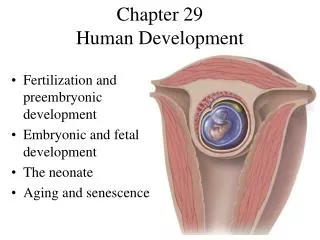
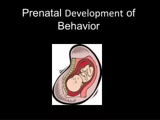
Process of Development • The vertebrate embryo develops in three stages • Cleavage • A hollow ball of cell forms • Gastrulation • Cells move to the interior, forming the primary tissues • Neurulation • The organs of the body form
Cleavage: From Zygote to Blastocyst • The first cleavage produces two daughter cells called blastomeres • Morula – the 16 or more cell stage (72 hours old) • By the fourth or fifth day the preembryo consists of 100 or so cells (blastocyst) • Blastocyst – a fluid-filled hollow sphere composed of: • A single layer of trophoblasts • A fluid-filled cavity, the blastocoel • An inner cell mass • Trophoblasts take part in placenta formation • The inner cell mass becomes the embryonic disc (the embryo)
Extraembryonic Membranes • The embryo reaches the uterus on day 6 • It penetrates the endometrial lining & initiates membrane formation • Amnion • Encloses embryo • Chorion • Forms from the trophoblast • Interacts with uterine tissue to form the placenta • Chorion • Yolk sac • Allantois • Amnion
Certain groups of cells move inwards from the inner cell mass at about 10-11 days after fertilization This process of gastrulation results in the three primary germ layers Endoderm Ectoderm Mesoderm Gastrulation: Onset of Developmental Change
Neurulation: Determination of Body Architecture • In the third week, the three primary germ layers begin development into body tissues and organs • First, the neural tube develops from the ectoderm • The notochord develops from the mesoderm • The gut develops from the endoderm • On either side of the notochord blocks of tissue (somites) form • These give rise to muscles, vertebrae and connective tissues developing notochord • By the end of the third week, the embryo is about 2 mm (< 0.1 inches) long
Fetal Development: 4th Week Fourth week • Formation of body organs, or organogenesis • Critical time in development • Alcohol use may cause fetal alcohol syndrome • Embryo reaches about 5 mm
Fetal Development: 2nd Month Second month • Great changes in morphology occur • Limbs assume adult shape • Major internal organs are evident • Embryo reaches about 25 mm
Fetal Development: 3rd Month Third month • Development is essentially complete except for lungs and brain • Developing human is now called a fetus • It carries out primitive reflexes like sucking
Fetal Development: 2nd Trimester Second trimester • A time of growth • Bone formation occurs • Hair and body are covered with fine hair called lanugo • By the end of the 6th month, the fetus is 30 cm (1 foot) long
Fetal Development: 3rd Trimester Third trimester • Pace of growth accelerates • Weight of fetus more than doubles as nutrients are still provided by mother’s blood via the placenta • Most major nerve tracts are formed in the brain
Postnatal Development • Babies typically double birth weight within a few months • Different body parts grow at different rates • Allometric growth • Nerve cells produced at an average rate of > 250,000 per minute • At 6 months, neuron production ceases permanently
Circulation in Fetus and Newborn • By the end of the 3rd week: • The embryo has a system of paired vessels • The vessels forming the heart have fused • Unique vascular modifications seen in prenatal development include umbilical arteries and veins, and three vascular shunts (occluded at birth) • Ductus venosus – venous shunt that bypasses the liver • Foramen ovale – opening in the interatrial septa to bypass pulmonary circulation • Ductus arteriosus – transfers blood from the right ventricle to the aorta
Effects of Pregnancy: Anatomical Changes • Chadwick’s sign – the vagina develops a purplish hue • Breasts enlarge and their areolae darken • The uterus expands, occupying most of the abdominal cavity • Lordosis is common due to the change of the body’s center of gravity • Relaxin causes pelvic ligaments and the pubic symphysis to relax • Typical weight gain is about 29 pounds
Effects of Pregnancy: Metabolic Changes • The placenta secretes human placental lactogen (hPL), also called human chorionic somatomammotropin (hCS), which: • Stimulates the maturation of the breasts • Promotes growth of the fetus • Exerts a maternal glucose-sparing effect • Human chorionic thyrotropin (hCT) increases maternal metabolism • Parathyroid hormone levels are high, ensuring a positive calcium balance
Effects of Pregnancy: Physiological Changes • GI tract – morning sickness occurs due to elevated levels of estrogen and progesterone • Urinary system – urine production increases to handle the additional fetal wastes • Respiratory system – edematous and nasal congestion may occur • Dyspnea (difficult breathing) may develop late in pregnancy • Cardiovascular system – blood volume increases 25-40% • Venous pressure from lower limbs is impaired, resulting in varicose veins
Parturition: Initiation of Labor • Estrogen reaches a peak during the last weeks of pregnancy causing myometrial weakness and irritability • Weak Braxton Hicks contractions may take place • As birth nears, oxytocin and prostaglandins cause uterine contractions • Emotional and physical stress: • Activates the hypothalamus • Sets up a positive feedback mechanism, releasing more oxytocin
Stages of Labor: Dilation Stage • From the onset of labor until the cervix is fully dilated (10 cm) • Initial contractions are 15–30 minutes apart and 10–30 seconds in duration • The cervix effaces and dilates • The amnion ruptures, releasing amniotic fluid (breaking of the water) • Engagement occurs as the infant’s head enters the true pelvis
Stages of Labor: Expulsion Stage • From full dilation to delivery of the infant • Strong contractions occur every 2–3 minutes and last about 1 minute • The urge to push increases in labor without local anesthesia • Crowning occurs when the largest dimension of the head is distending the vulva
Stages of Labor: Expulsion Stage • The delivery of the placenta is accomplished within 30 minutes of birth • Afterbirth – the placenta and its attached fetal membranes • All placenta fragments must be removed to prevent postpartum bleeding
Apgar Score • At 1-5 minutes after birth, the infant’s physical status is assessed based on five signs: heart rate, respiration, color, muscle tone, and reflexes • Each observation is given a score of 0 to 2 • Apgar score = the total score of the above assessments • 8-10 indicates a healthy baby • Lower scores reveal problems