Download
1 / 45
510 likes | 929 Vues
Caenorhabditis elegans (C. elegans) An elegant worm. Why study worms?. Sydney Brenner.

E N D
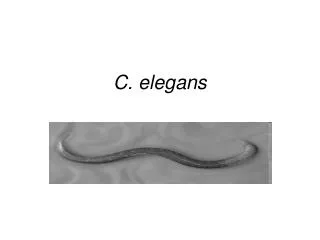
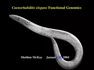
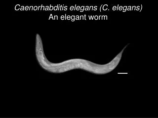
Caenorhabditis elegans (C. elegans) An elegant worm
Why study worms? Sydney Brenner “Thus we want a multicellular organism which has a short life cycle, can be easily cultivated, and is small enough to be handled in large numbers, like a micro-organism. It should have relatively few cells, so that exhaustive studies of lineage and patterns can be made, and should be amenable to genetic analysis.” --Excerpts from Proposal to the Medical Research Council, 1963
C. elegans: the chosen one! Easily cultivated: can grow thousands on a petri dish, feed on non-hazardous bacteria, and cheap to maintain Short generation time: 3 days Small: 1 mm (about the size of a pinhead) Few cells: The adult has 959 hermaphrodrodite (XX) or 1031 (XO) cells Amenable to genetic analysis: maintained as hermaphrodites, but males exist for genetic studies,The genome is small- 100 Mb Transparency: allows for development to be analyzed from a single cell and all cells to be lineage Photo credit: Ian D. Chin-Sang (Queen's University, Kingston, ON, Canada).
Life cycle of C. elegans Photo credit: http://www.scq.ubc.ca/genetic-studies-of-aging-and-longevity-in-model-organisms/
Anatomy of C. elegans Anus Pharynx Rectum Intestine (yellow) Epidermis Gonad (pink) Vulva head ~1 mm tail anterior posterior Fig. 8.43
Hermaphrodites do it by themselves Hermaphrodite (XX) Males (X0) Photo credit: http://homepages.ucalgary.ca/~dhansen/worms.gif
The C. elegans gonad: an extremely efficient reproductive system Fig. 8.42
Within this lineage is the secret of embryonic development John Sulston
An entire C. elegans hermaphrodite worm consists of exactly 959 cells EVERY SINGLE TIME, allowing one to follow the cell lineage. Learn to read a lineage diagram! Branching = Increasing cell division age of worm embryo 1st stage larva 2nd stage larva Line ending = differentiated cell = Cell death
P0 zygote 2 cell stage 4 cell stage 8 cell stage Cleavage Events Lineage
Most lineages consist of multiple tissue types but the P4, E and D cells gives rise to a single tissue type Fig. 8.43
Question: 1.) How many cell divisions took place in the wildtype lineage? ____ 2.) In wild-type, how many total descendants will cell A have? ____ 3.) How many differentiated cells from the wild-type lineage will be a part of the adult worm? ____ 4.) What is the best description of the defect in mutant 1?
How are the invariant lineages established? ie. How do cells know who they are and what they are doing? • Control of apoptosis • Partitioning of cytoplasmic determinants • Timing of developmental events • Cell-Cell interactions
Even cell death is programmed into the lineage C. elegans was used to identify the machinery that regulates programmed cell death in vertebrates
The Nobel Prize in Physiology or Medicine 2002 "for their discoveries concerning ’ genetic regulation of organ development and programmed cell death'" Sidney Brenner H. Robert Horvitz John Sulston
Partitioning of cytoplasmic determinants P-granules (green) are cytoplasmic determinants that are formed from ribonucleoprotein complexes that specify the germ cells blue nuclei green P-granules P3 P0 P granules are asymmetrically segregated into one cell, the P4 cell, which will give rise to the germline P1 AB P4
PARtition mutants (PAR) disrupt the asymmetric distribution of p-granlues Photo credit: http://mbg.cornell.edu/cals/mbg/research/kemphues-lab/images/par_phenotypes.gif
Timing of developmental events Lof= loss of function, gene function is disrupted lin-4 (lof) lin-14 (lof) wildtype Moss E. 2007. Current Biology, R425. Lin-14 is required for the timing of cell division in the L1 stage. Lin-4 regulates transition from L1 to L2 stage. .
lin-4 (lof) lin-14 (lof) wildtype Graph of LIN-14 and LIN-4 levels in a wildtype embryo LIN-4 Levels LIN-14 Time L1 L2 L3 L4 Adult
If you have a mutation that results in an INCREASED level of LIN-14 (gain of function) which lineage would you expect lin-4 (lof) lin-14 (lof) wildtype Graph of LIN-14 and LIN-4 levels in a wildtype embryo LIN-4 Levels LIN-14 Time L1 L2 L3 L4 Adult
lin-4 does not encode a protein—what???? It encodes for a microRNA lin-4 lin-4 lin-14 lin-4 lin-14 Translation blocked!
Cell-Cell Interactions: the P2 impact! Glp-1/Notch receptor Apx-1/Delta-like ligand mom-2/ Wnt ligand mom-5/ Wnt receptor Signal from P2 cell required to induce EMS cell to produce E cell which forms the gut (see p. 248)
How to cell interactions relate to the formation of an organ? Vulva formation!
Getting the terminology down: C. elegans Vulva Early larval stage Figure 6.27 Anchor cell (AC) Gonad VPCs AC Basement membrane Gonad P3.p-P8.p are the Vulva Precursor Cells (VPCs) Later larval stage P5.p,P6.p and P7.p lineages make the vulva 3° 2° 2° 1° 3° P3.p,P4.p and P8.p lineages non-vulval P3.p P4.p P5.p P6.p P7.p P8.p
Inductive and lateral signals induce the vulva Anchor cell gonad P4 P7 P8 P3 P5 P6 VPCs VPCs after induction 1° 2° 3° 3° 3° 2° The primary and secondary cells form the vulva
How’d you know that? Cell ablation studies helped identify key players in vulva formation Lecture notes: experiment 1
If anchor cell signaling is disrupted, all VPCs cells adopt a non-vulva fate anchor cell gonad 3° cell 3° cell 3° cell 3° cell 3° cell 3° cell no vulva
Early stage The VPCs have multipotential Anchor cell gonad P8 P6 P3 P7 P5 P4 VPCs Later stage Anchor cell gonad 3° 3° 2° 3° 2° 1° What is causing the VPCs to be different?
Let’s do an experiment: what happens when the P6.p cell is ablated? Anchor cell gonad P8 P3 P7 P5 P4 VPCs A P6 B C 3° 3° 3° 3° 3° 3° 3° 2° 3° 2° 2° 2° 3° 3° 3° 3° 2° 3° 1° 2° 1° Lecture notes: experiment 2
What genes specify the VPC cell fate? Looked for mutants that disrupted vulva formation 1) No vulva: worms hatch inside (yuck!!) 1) Too many vulvas Lecture notes: experiment 3
Inductive and lateral signals induce the vulva Anchor cell gonad P4 P7 P8 P3 P5 P6 VPCs VPCs after induction 1° 2° 3° 3° 3° 2° The primary and secondary cells form the vulva
The vulvaless mutations helped define the Ras pathway Lin-3/Epidermal Growth Factor (EGF) Let-23/EGF Receptor Let-60/RAS Sem-5/GRB2 Lin-45/RAF P6.p becomes the primary cell!
The Ras pathway is abnormally activated in many human tumors eg: pancreatic cancer, colorectal cancer, lung adenocarcinoma, gall bladder cancer, bile duct cancer and thyroid cancer Another representation of the RAS pathway (VPC cells) LIN-3 signal
The Ras mutation is so prevalent that kits are available to test of mutations that are linked to cancer
A signal from P6.p actives notch (lin-12) in P5.p and P7.p Figure 6.27
The transmembrane receptor is the Lin-12 protein, a receptor protein related to Notch “ Primary cell” “ Secondary cells” Both membrane and receptor are membrane bound!
Generation of Different Cell Types From Equivalent Cells in C. elegans:Initial specification of the Anchor Cell also requires Notch The signal: lag-2 (delta) The receptor: lin-12 (notch) Figure 6.28
Does the Notch pathway remind you of anything you learned earlier? No notch=neural!
The story of epidermal vs. neuronal fate in Drosophila If signal is missing... Some cells become neuroblasts and signal their neighbors to remain epidermis all cells eventually ingress and become neuroblasts Nervous system Extra nervous system Epidermis No epidermis!