Peripheral contractile mediators
Peripheral contractile mediators. 1-Neural factors : NA , NPY (Neuropeptid Y .) 2- Endothelial factors : ET 1 , PGF 2 α ,Thromboxane A 2 & Angiotensin II(synthetised by penile tissue). Sm Contraction. NA, ET 1 , PGF 2 α. Via their receptors.
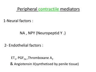

Peripheral contractile mediators
E N D
Presentation Transcript
Peripheral contractile mediators 1-Neural factors : NA , NPY (Neuropeptid Y .) 2- Endothelial factors : ET1, PGF2α ,Thromboxane A2 & Angiotensin II(synthetised by penile tissue)
Sm Contraction NA, ET1, PGF2α Via their receptors • Stimulate G Protein Receptor • Phospholipase C • ( Intracellular Ca++) Activte G Protein Coupled Receptor Rho/RhoKinase ( Calcium Sensitivity pathway)
NA,ET-1,PGF2A via their Rs PGRs (protein G receptor). • PG activate PLc (phospholipase C enzyme) • Phosphatidylinstol biphosphate (PIP ) by the effect of PLc Diacylglycerol PKC IP3 (inositol triphosphate)
Intracellular Ca++ • At the level of Cell Membrane • PKC Ca++ influx • At the Sarcoplasmic reticulum • IP3 release of Ca++ from SR So the intracellular Ca++
intracellular Ca++ is transitory. • Then the Sm maintains contraction after the intracellular Ca++ returns to the basal level. • So the phasic contraction of P.Sm is regulated by intracellular Ca++
Cont. (Calcium Sensitivity Pathway) • NA, ET1, PGF2α +GP coupled receptors activate RhoA/RhoKinase pathway. Ca++ sensitivity ( not the free intracellular ca++).
NA, ET1, PGF2α + G-P coupled rec RhoA RhoK MLC phosphatase MLC kinase Phosphorylated MLC SM Contraction
Small GTP-binding proteins (G proteins), include Rho, Ras, Rab, Sarl/Arf, and Ran families. • They play a substantial role in intracellular signaling pathways. • At least 10 members of the Rho family are present in mammals, including Rho (isoforms A to E, and G), Rac (isoforms 1 to 3), Cdc42, and TC10. • Rho is known to modulate Ca2+-sensitization of vascular smooth muscle cells and inhibit myosin phosphatase activity. • Rho-Kinase is one of the effectors of the small GTP-binding protein Rho.
At cellular level, Rho/Rho-kinase pathway plays an important role in: - vascular smooth muscle cell (VSMC) contraction. -Actin cytoskeleton organization. -Cell adhesion and motility. -Cytokinesis. -Gene expressions. • At molecular level, Rho-kinase upregulates various molecules that accelerate inflammation/oxidative stress, thrombus formation, and fibrosis, whereas it downregulates endothelial nitric oxide synthase.
RhoA, a member of the Ras low molecular weight of GTP-binding proteins, mediates agonist-induced activation of Rho-kinase. • The exchange of GDP for GTP on RhoA and translocation of RhoA from the cytosol to the membrane are markers of its activation and enable the downstream stimulation of various effectors such as Rho-kinase. • Both human endothelial cells and human corpus cavernosum smooth muscle cells express RhoA and Rho-kinase.
NO inhibits Rho-kinase activity, and the endogenous NO-mediated vasodilation may occur through the inhibition of Rho-kinase vasoconstrictor activity. • The RhoA/Rho-kinase pathway plays an important role in suppression of eNOS gene expression and enzyme activity in human endothelial cells, which results in decreased endothelial-derived NO biosynthesis.
Rhoa/rho-kinase pathway • ROK (serine-threonine kinase) • ROK P-MLC S.Ms. Contraction What is (Y27632)? It is a selective inhibitor of ROK and has been shown to cause SM relaxation and erection
Contraction • Thephasic contraction of penile Sm. is regulated by free intracellular Ca++ • whiletonic contractionis governed by Ca++ sensitizing pathway. • Thistonic contraction is responsible for the physiological flaccid statefor the majority of time.
The maintenance of penile detumescence depends on: 1- Anti-erectile RhoA/Rho-Kinase Ca++ sensitivity pathway. 2- The PDE5 degradation of CGMP