Download
1 / 63
630 likes | 645 Vues
This topic explores the functions and regulation of the male reproductive system, including spermatogenesis, the role of hormones, semen composition, and the control of testosterone secretion.

E N D
Objectives At the end of the topic, the students should be able to : • Explain the functions of the testis • Discuss spermatogenesis • Describe the characteristics of a normal sperm • Explain the role of FSH and inhibin on spermatogenesis • Discuss the contents of semen and its abnormalities • Explain the functions of testosterone • Explain the control of secretion of testosterone
Main points in male reproductive physiology • Importance of Leydig cells and testosterone in puberty • Role of Sertoli cells,testosterone, DHT, and estrogen in spermatogenesis • How testosterone and inhibin functions in the adult feedback regulation of the Hypothalamic/ Pituitary/ Gonadal Axis
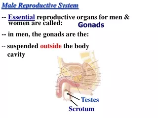
INTRODUCTION • A pair of testes • A pair of accessory glands • Ductal system • Copulatory organ
FUNCTIONS • Production of spermatozoa after puberty for fertilization with the ovum from the female • Coitus process • Produce androgens
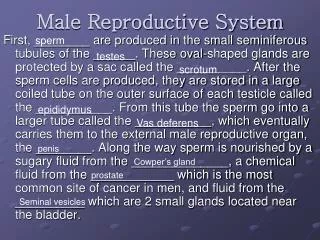
Testis • Originates from indifferent gonads during the embryonic phase • Contains lobules (200-300) separated by septum • Each lobule has 4 seminiferous tubules • Blood supply is from testicular artery and blood drainage is through the Pampiniform plexus into the testicular vein • Seminiferous epithelium contains Sertoli cells (Sustentacular cells) and Germ cells • Leydig cells exist in between tubules • Produce spermatozoa and androgens • Divided into two compartments; extratubular and intratubular
Histology of the testis ETC ITC Spermatogonia Leydig cells SCN Lumen
Compartments • Extratubular – vascular and interstitial divisions (inclusive of lymphatic channels and Leydig cells) • Intratubular – basal and adluminal divisions located in the seminiferous tubules
Blood Testis Barrier • Formed before spermatogenesis commences • Formed by Sertoli cells • Functions : • Stop intratubular spermatozoa from entering systemic and lymphatic circulation The blood testes barrier is important since sperm (with their unique surface antigens) elicit an immune response if detected by the immune cells in the man’s blood, and the antibodies that are formed against sperm are designed to immobilize and destroy them
A number of events can disrupt the blood testes barrier and allow the immune system to become activated against the sperm. These include: • trauma to the testes • torsion (twisting) of the testes • a history of a vasectomy and reversal • any other surgery within the scrotum • infection within the testes
Allergic orchitis Inflammation of the testis due to antisperm antibodies
Ensure the intratubular chemical composition is different from the intertubular chemical composition (blood, interstitial fluid and lymph) As spermatogenesis must occur in a controlled microenvironment, any changes in the chemical composition will affect normal spermatogenesis
Functions of Sertoli cells • Sertoli cells are joined by tight junctions that provides barrier to chemicals (Blood testis barrier) • Nourish developing sperm • Secrete luminal fluid including androgen binding proteins (ABP) • Phagocytize defective sperm • Mediate testosterone and FSH effects on spermatogenesis • Secrete inhibin which inhibits FSH secretion • Influence Leydig cells via paracrine secretions • Embryonic secretion of Mullerian Inhibiting Substance (MIS) that ensures male phenotype
Spermatogenesis • Process for formation of spermatozoa • The seminiferous tubules produce haploid cells (n) • Involves several steps including mitosis and meiosis • Takes about 64 days in human • Consists of three stages: • Spermacytogenesis – formation of spermatid from spermatogonia • Spermiogenesis – formation from spermatid to spermatozoa • Spermiation – the release of spermatozoa into the epididymis
Spermacytogenesis • Mitosis stage • spermatogonia type Ad give rise to a pair of spermatogonia type Ad or Ap • spermatogonia type Ap give rise to a pair of spermatognia type Ap or B • mitosis of the germ cells occurs in the basal compartment • incomplete cytokenesis of committed cells (type Ap) results in the cells being linked by cytoplasmic bridges until spermatozoa is form • this linkage results in a synchronous development of the cells within a given region of the tubule
Types of cells • germ cells - - spermatogonia (types A and B): • spermatogonia type A - for spermatogenic lineage (type A dark (Ad) or A pale (Ap), type Ad are true stem cells) • spermatogonia type B - progenitor cell for primary spermatocyte • primary spermatocyte (46 chromosomes, 4N DNA): • first meiotic division from these cells result in secondary spermatocyte (23 chromosomes, 2N DNA) • second meiotic division from these cells results in spermatids • spermatids (23 chromosomes, 1N DNA) – undergoes modifications in many parts and gives rise to spermatozoa • spermatozoa, within the seminiferous tubules (and distally) the flagella is not motile
Spermiogenesis • spermatid phase: spermatids develop into spermatozoa in 4 stages: • Golgi phase - development of the acrosomal granule from the Golgi complex forming the acrosomal vesicle at the nascent apex (anterior) of cell • positioning of centrioles at the nascent base of flagella • initiation of formation of axonemal complex from one of the centrioles • Cap phase • development of acrosomal cap over nucleus and condensation of chromatin • development of flagellum from axonemal complex • acrosome contains hydrolases (proteases, hyaluronidase, neuramidase, acid phosphatase) important in penetration of the oocyte membrane during fertilization
Spermiogenesis • Acrosome phase - spermatid re-orients so tail (flagellum) projects into the lumen of the tubule and the acrosome towards the base of the epithelium • further condensation of chromatin • flattening and elongating of nucleus at the anterior of the cell • movement of cytoplasm to the posterior of cell • further development of flagellum • linkage of flagellum to nucleus via the connecting piece developed from a centriole • Maturation phase - residual body of cytoplasm shed (cell linkages lost) and the cells released by Sertoli cell into lumen of tubule
Spermiation • released spermatid moved with fluid via peristaltic action of myoid cells transporting it to the straight tubule • sperm cannot move yet • will develop motility in epididymis
1 germ cell produces 64 spermatozoa • Process takes ~2 months (56 – 64 days ) to complete • 100 million sperm produced each day • Not all are normal i.e., abnormal % increases with alcohol, heat, cigarettes, drugs
Temperature sensitivity… • Spermatogenesis is temperature sensitive, optimal 34o C • Achieved by the descent of the testes out of the abdomen • Arteries and veins supplying the testes intertwine, efficiently exchanging heat (from artery to veins) to further cool testes
Scrotum • Originates from labioscrotal swellings and urethral fold • Pouch that houses the testes • Main function is to provide an environment which is 1-80F lower than the body temperature and also to control testicular temperature • Testicular temperature needs to be controlled for spermatogenesis to occur normally
Control and regulation of testicular temperature (1) • Two muscle system – cremaster external and tunica dartos • Cremaster external muscle passes along inguinal canal and attaches to tunica vaginalis • Pulls tunica vaginalis as it contracts when the environmental temperature drops • Tunica dartos muscle attaches to scrotal skin and forms a septum separating the scrotum - Pulls scrotal skin as it contracts when the environmental temperature drops
Control and Regulation of testicular temperature (2) • Pampiniform plexus – consists of convoluted veins and arteries which follow the spermatid cord into the inguinal canal. Arterial branch comes from spermatid artery and venous part enters the spermatid vein - controls temperature by dissipating heat from the aortal blood through the convolutions before reaching the testis
The testes is located in the abdominal cavity during the fetal stage • Only descents into scrotum at 7 months of pregnancy • Sometimes do not descend and when born, two conditions may occur i.e., cryptochidism or monochidism • Crypotochidism – cryptochids are males with both testes in inguinal canal/abdominal cavity • usually sterile • may either be hereditary or due to lack of hormones • undescended testes are associated with reduced fertility, increased risk of testicular cancer and psychological problems when the boy is grown • undescended testes are also more susceptible to testicular torsion, infarction and inguinal hernias
Monochidism – monochids are males with only one testis descended into scrotum, the other remained in inguinal canal/abdominal cavity - usually fertile as one testis still functioning normally therefore spermatogenesis is not impaired • Approximately 3% of full-term and 30% of premature infant boys are born with at least one undescended testis, making monochidism/cryptorchidism the most common birth defect of male genitalia • However, most testes descend by the first year of life (the majority within three months), making the true incidence of cryptorchidism around 1% overall.
Ductal System • Originates from Wolffian ducts (mesonephric kidney) • Mullerian ducts = rudiments in prostate gland (prostatic utricle/uterus masculinus) – non-functional but can grow when there is estrogen influence causing prostate cancer • Mesonephric tubules = vasa efferentia • Mesonephric ducts = epididymis, vas deferens and seminal vesicles • Urogenital sinus = prostatic, cavernous and membranous urethra, prostate glands and Bulbo-urethral gland (Cowper’s gland) • Rete testis in the testis = efferentia ducts and then becomes epididymis and vas deferens
Epididymis • Originates from mesonephric ducts • Divided into caput (head), corpus (body) and cauda (tail) • Extra fluid from sperm is reabsorbed to concentrate spermatozoa 100X • Presence of high concentrations of testosterone/ DHT in the tubule causes epididymis to secrete a motility- inducing protein that binds to the cell membrane of sperm cells • Secrete mucoproteins/glycoproteins that coats the head of the sperm • Also secretes carnitine, glycerolphosphorycholine, fructose and glycoproteins • Transit time for spermatozoa to attain maturation and the ability to move/motility is about 6 – 12 days • Spermatozoa can move forward and has the ability to fertilize ovum once has entered cauda epididymis • Morphological and biochemical changes also occur
Caput Corpus Cauda
Vas deferens • Developed from Wolffian ducts • Vas deferens have a lot of muscle layers (inner and outer longitudinal muscle layer with circular layers in between this muscles – layers are important for sperm motility • Vas deferens form the ampulla near the bladder • Sperm enters vas deferens from epididymis • Acts as a reservoir to store spermatozoa • Also acts as a conduit between testes and urethra • Mature sperm stored in the Vas Deferens and can remain viable for up to 3 months • If no ejaculation occurs, sperm will dribble into terminal ampulla into urethra
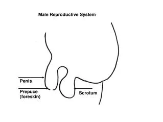
Copulatory organ - penis • Originates from genital tubercle • Provides an outlet for both urine and the copulatory ejaculate (spermatozoa and seminal plasma) • Histology/anatomy of penis varies from species to species and from region to region within the same species • Body of the penis consists of: • the urethra • erectile tissue (corpora cavernosa penis and corpora cavernosum urethra), cavernous bodies act as erectile tissues where it can be engorged with blood to erect the penis • touch and pressure receptors (Pacinian corpuscles) • a dense connective tissue capsule (tunica albuginea • tip of penis is called glans penis and in humans it is mushroom-shaped
Accessory glands • A pair of seminal vesicles, prostate glands and bulbo-urethral glands (Cowper’s glands) • Originates from urogenital sinus and mesonephric ducts • Produces seminal plasma which is the fluid component of semen
seminal vesicle empties contents into ampulla • tubuloalveolar gland • yellowish secretory product and contains fructose, citrate, ascorbic acid, inositol, prostaglandins and proteins • fructose is an important energy source for spermatozoa • 70% of ejaculate (semen) derives from the seminal vesicles • secretory activity stimulated by testosterone • prostrate gland empties contents into the prostatic urethra • prostrate is a branched tubuloalveolar gland • secretions include acid phosphatase, amylase, and fibrinolysin • gland wraps around prostatic urethra and empties into it • glycoprotein deposits develop and can become calcified, these are called prostatic concretions or corpora amylacea and are a characteristic feature of the adult prostrate • secretory activity stimulated by testosterone • hyperplasia occurs within the gland with increasing age, gradually decreasing the diameter of the urethra and slowing urine voiding
bulbourethral (Cowper’s) gland empty into the post-prostatic urethra • tubuloalveolar glands that secrete a lubricating mucous that contains galactose, galactosamine, galacturonic acid, sialic acid, and methylpentose • secretions precede other ejaculatory products and release involves the oxytocin axis • secretory activity of epithelia stimulated by testosterone
Seminal plasma Three functions: • as a media that provides the suspension/vehicle and also activation to spermatozoa • provides electrolytes, nitrogen, citric acid, fructose etc for nutrition • provides an alkaline pH to semen to combat acidity of vagina
Spermatozoa • A very simple cell but highly adapted for reaching and penetrating the ovum • Basically have head, midpiece and tail • Different sizes in different species • Head shape may be paddle (human, rabbit, bull, ram), cylindrical (cockerel) or hooked (rodent species) • Acrosome in the head contains hydrolytic enzymes for digesting cells around ovum; can also digests sperm upon death • Carries the necessary package for fertilization (enzymes and chromosomes) Needs to travel light as main function is for fertilization • Tail contains mitochondria that provides ATP for energy for increased motility to reach ovum before sperm dies • Tail have 9 + 2 doublets arrangement called axoneme • Has no energy reserves, hence has a limited life span once released (48 – 72 hrs) • Dies by degeneration (hydrolytic enzymes in acrosome destroy sperm upon death)