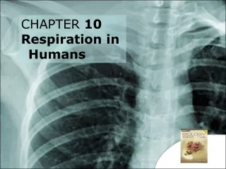
Chapter 10 Respiration During Exercise
Chapter 10 Respiration During Exercise. EXERCISE PHYSIOLOGY Theory and Application to Fitness and Performance, 5 th edition Scott K. Powers & Edward T. Howley. Introduction. The Respiratory System Provides a means of gas exchange between the environment and the body

Chapter 10 Respiration During Exercise
E N D
Presentation Transcript
Chapter 10Respiration During Exercise EXERCISE PHYSIOLOGY Theory and Application to Fitness and Performance, 5th edition Scott K. Powers & Edward T. Howley
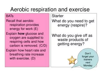
Introduction • The Respiratory System • Provides a means of gas exchange between the environment and the body • Plays a role in the regulation of acid-base balance during exercise
Objectives • Explain the principle physiological function of the pulmonary system • Outline the major anatomical components of the respiratory system • List major muscles involved in inspiration and expiration, at rest and during exercise • Discuss the importance of matching blood flow to alveolar ventilation in the lung • Explain how gases are transported across the blood-gas interface in the lung
Objectives • Discuss the major transportation modes of O2 and CO2 in the blood • Discuss the effects of temp, pH, and levels of 2-3 DPG on the oxygen-hemoglobin dissociation curve • Describe the ventilatory response to constant load, steady-state exercise
Objectives • Describe the ventilatory response to incremental exercise • Identify the location and function of chemoreceptors and mechanoreceptors that are thought to play a role in the regulation of breathing • Discuss the neural-humoral theory of respiratory control during exercise
Respiration • Pulmonary respiration • Ventilation (breathing) and the exchange of gases (O2 and CO2) in the lungs • Cellular respiration • Relates to O2 utilization and CO2 production by the tissues • This chapter is concerned with pulmonary respiration, and “respiration” will be used to mean such
Function of the Lungs • Primary purpose is to provide a means of gas exchange between the external environment and the body • Ventilation refers to the mechanical process of moving air into and out of lungs • Diffusion is the random movement of molecules from an area of high concentration to an area of lower concentration
Conducting zone Conducts air to respiratory zone Humidifies, warms, and filters air Components: Trachea Bronchial tree Bronchioles Respiratory zone Exchange of gases between air and blood Components: Respiratory bronchioles Alveolar sacs Conducting and Respiratory Zones
Conducting & Respiratory Zones Fig 10.2
Pathway of Air to Alveoli Fig 10.4
Mechanics of Breathing • Inspiration • Diaphragm pushes downward, lowering intrapulmonary pressure • Expiration • Diaphragm relaxes, raising intrapulmonary pressure • Resistance to airflow • Largely determined by airway diameter
Muscles of Respiration Fig 10.7
Pulmonary Ventilation (V) • The amount of air moved in or out of the lungs per minute • Product of tidal volume (VT) and breathing frequency (f) V = VTx f
Pulmonary Ventilation (V) • Dead-space ventilation (VD) • “Unused” ventilation • Does not participate in gas exchange • Anatomical dead space: conducting zone • Physiological dead space: disease • Alveolar ventilation (VA) • Volume of inspired gas that reaches the respiratory zone V = VA + VD
Pulmonary Volumes and Capacities • Measured by spirometry • Vital capacity (VC) • Maximum amount of air that can be expired following a maximum inspiration • Residual volume (RV) • Air remaining in the lungs after a maximum expiration • Total lung capacity (TLC) • Sum of VC and RV
Pulmonary Volumes and Capacities Fig 10.9
PO2 = 0.2093 x 760 = 159 mmHg Partial Pressure of GasesDalton’s Law • The total pressure of a gas mixture is equal to the sum of the pressure that each gas would exert independently • The partial pressure of oxygen (PO2) • Air is 20.93% oxygen • Expressed as a fraction: 0.2093 • Total pressure of air = 760 mmHg
A V gas = x D x (P1-P2) T Diffusion of GasesFick’s law of diffusion • The rate of gas transfer (V gas) is proportional to the tissue area, the diffusion coefficient of the gas, and the difference in the partial pressure of the gas on the two sides of the tissue, and inversely proportional to the thickness. V gas = rate of diffusion D = diffusion coefficient of gas A = tissue area P1-P2 = difference in partial pressure T = tissue thickness
Partial Pressure and Gas Exchange Fig 10.10
Blood Flow to the Lung • Pulmonary circuit • Same rate of flow as systemic circuit • Lower pressure Fig 10.11
Blood Flow to the Lung • When standing, most of the blood flow is to the base of the lung • Due to gravitational force Fig 10.12
Ventilation-Perfusion Relationships • Ventilation/perfusion ratio • Indicates matching of blood flow to ventilation • Ideal: ~1.0 • Base • Overperfused (ratio <1.0) • Apex • Underperfused (ratio >1.0)
Ventilation/Perfusion Ratios Fig 10.13
O2 Transport in the Blood • Approximately 99% of O2 is transported in the blood bound to hemoglobin (Hb) • Oxyhemoglobin: O2 bound to Hb • Deoxyhemoglobin: O2 not bound to Hb • Amount of O2 that can be transported per unit volume of blood in dependent on the concentration of hemoglobin
Oxyhemoglobin Dissociation Curve Fig 10.14
O2-Hb Dissociation Curve: Effect of pH • Blood pH declines during heavy exercise • Results in a “rightward” shift of the curve • Bohr effect • Favors “offloading” of O2 to the tissues Fig 10.15
O2-Hb Dissociation Curve: Effect of Temperature • Increased blood temperature results in a weaker Hb-O2 bond • Rightward shift of curve • Easier “offloading” of O2 at tissues Fig 10.16
O2-Hb Dissociation Curve: 2-3 DPG • RBC must rely on anaerobic glycolysis to meet the cell’s energy demands • A by-product is 2-3 DPG, which can combine with hemoglobin and reduce hemoglobin’s affinity of O2 • 2-3 DPG increase during exposure to altitude • At sea level, right shift of curve not to changes in 2-3 DPG, but to degree of acidosis and blood temperature
O2 Transport in Muscle • Myoglobin (Mb) shuttles O2 from the cell membrane to the mitochondria • Higher affinity for O2 than hemoglobin • Even at low PO2 • Allows Mb to store O2
CO2 Transport in Blood • Dissolved in plasma (10%) • Bound to Hb (20%) • Bicarbonate (70%) • CO2 + H2O H2CO3 H+ + HCO3- • Also important for buffering H+
CO2 Transport in Blood Fig 10.18
Release of CO2 From Blood Fig 10.19
Rest-to-Work Transitions • Initially, ventilation increases rapidly • Then, a slower rise toward steady-state • PO2 and PCO2 are maintained Fig 10.20
Exercise in a Hot Environment • During prolonged submaximal exercise: • Ventilation tends to drift upward • Little change in PCO2 • Higher ventilation not due to increased PCO2 Fig 10.21
Incremental Exercise • Linear increase in ventilation • Up to ~50-75% VO2max • Exponential increase beyond this point • Ventilatory threshold (Tvent) • Inflection point where VE increases exponentially
Ventilatory Response to Exercise:Trained vs. Untrained • In the trained runner, • decrease in arterial PO2 near exhaustion • pH maintained at a higher work rate • Tvent occurs at a higher work rate Fig 10.22
Ventilatory Response to Exercise:Trained vs. Untrained Fig 10.22
Exercise-Induced Hypoxemia • 1980s: 40-50% of elite male endurance athletes were capable of developing • 1990s: 25-51% of elite female endurance athletes were also capable of developing • Causes: • Ventilation-perfusion mismatch • Diffusion limitations due to reduce time of RBC in pulmonary capillaries due to high cardiac outputs
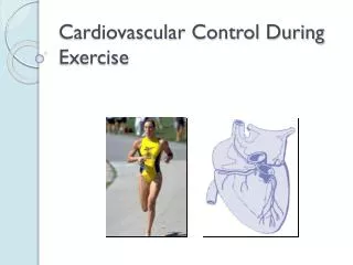
Control of Ventilation • Respiratory control center • Receives neural and humoral input • Feedback from muscles • CO2 level in the blood • Regulates respiratory rate Fig 10.23
Input to the Respiratory Control Centers • Humoral chemoreceptors • Central chemoreceptors • Located in the medulla • PCO2 and H+ concentration in cerebrospinal fluid • Peripheral chemoreceptors • Aortic and carotid bodies • PO2, PCO2, H+, and K+ in blood • Neural input • From motor cortex or skeletal muscle
Effect of Arterial PCO2on Ventilation Fig 10.24
Effect of Arterial PO2on Ventilation Fig 10.25
Ventilatory Control During Exercise • Submaximal exercise • Linear increase due to: • Central command • Humoral chemoreceptors • Neural feedback • Heavy exercise • Exponential rise above Tvent • Increasing blood H+
Effect of Training on Ventilation • Ventilation is lower at same work rate following training • May be due to lower blood lactic acid levels • Results in less feedback to stimulate breathing
Effects of Endurance Training on Ventilation During Exercise Fig 10.27