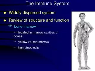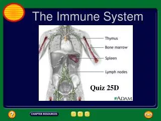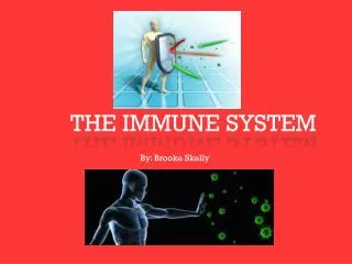
The Immune System
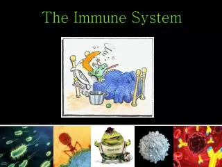
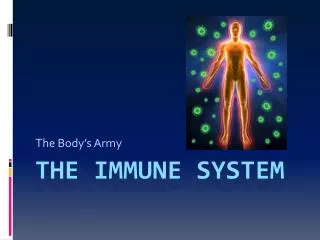
Fig. 43-1. The Immune System. 1.5 µm. Overview: Reconnaissance, Recognition, and Response. Barriers help an animal to defend itself from the many dangerous pathogens it may encounter The immune system recognizes foreign bodies and responds with the production of immune cells and proteins

The Immune System
E N D
Presentation Transcript
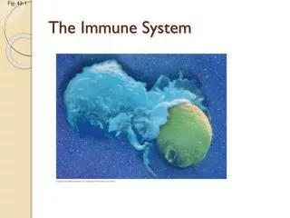
Fig. 43-1 The Immune System 1.5 µm
Overview: Reconnaissance, Recognition, and Response • Barriers help an animal to defend itself from the many dangerous pathogens it may encounter • The immune system recognizes foreign bodies and responds with the production of immune cells and proteins • Two major kinds of defense have evolved: innate immunity and acquired immunity
Innate immunity is present before any exposure to pathogens and is effective from the time of birth • It involves nonspecific responses to pathogens • Innate immunity consists of external barriers plus internal cellular and chemical defenses
Acquired immunity, or adaptive immunity, develops after exposure to agents such as microbes, toxins, or other foreign substances • It involves a very specific response to pathogens
Fig. 43-2 Pathogens (microorganisms and viruses) Barrier defenses: Skin Mucous membranes Secretions INNATE IMMUNITY • Recognition of traits shared by broad ranges of pathogens, using a small set of receptors Internal defenses: Phagocytic cells Antimicrobial proteins Inflammatory response Natural killer cells • Rapid response Humoral response: Antibodies defend against infection in body fluids. ACQUIRED IMMUNITY • Recognition of traits specific to particular pathogens, using a vast array of receptors Cell-mediated response: Cytotoxic lymphocytes defend against infection in body cells. • Slower response
In innate immunity, recognition and response rely on shared traits of pathogens • Both invertebrates and vertebrates depend on innate immunity to fight infection • Vertebrates also develop acquired immune defenses
Innate Immunity of Invertebrates • In insects, an exoskeleton made of chitin forms the first barrier to pathogens • The digestive system is protected by low pH and lysozyme, an enzyme that digests microbial cell walls • Hemocytes circulate within hemolymph and carry out phagocytosis, the ingestion and digestion of foreign substances including bacteria
Fig. 43-3 Microbes PHAGOCYTIC CELL Vacuole Lysosome containing enzymes
Innate Immunity of Vertebrates • The immune system of mammals is the best understood of the vertebrates • Innate defenses include barrier defenses, phagocytosis, antimicrobial peptides • Additional defenses are unique to vertebrates: the inflammatory response and natural killer cells
Barrier Defenses • Barrier defenses include the skin and mucous membranes of the respiratory, urinary, and reproductive tracts • Mucus traps and allows for the removal of microbes • Many body fluids including saliva, mucus, and tears are hostile to microbes (contain enzyme lysozyme) • The low pH of skin and the digestive system prevents growth of microbes
Cellular Innate Defenses • White blood cells (leukocytes) engulf pathogens in the body • Groups of pathogens are recognized by common receptors: lipopolysaccarides common to many bacteria, viral proteins or single-stranded DNA or RNA.
Fig. 43-6 EXTRACELLULAR FLUID Lipopolysaccharide Helper protein Flagellin TLR4 WHITE BLOOD CELL TLR5 VESICLE TLR9 CpG DNA Inflammatory responses TLR3 ds RNA
A white blood cell engulfs a microbe, then fuses with a lysosome to destroy the microbe • There are different types of phagocytic cells: • Neutrophils engulf and destroy microbes • Macrophages are part of the lymphatic system and are found throughout the body • Eosinophils discharge destructive enzymes • Dendritic cells stimulate development of acquired immunity
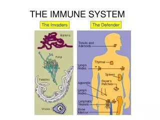
Fig. 43-7 Interstitial fluid Adenoid Tonsil Blood capillary Lymph nodes Spleen Lymphatic vessel Tissue cells Peyer’s patches (small intestine) Appendix Lymphatic vessels Lymph node Masses of defensive cells
Antimicrobial Peptides and Proteins • Peptides and proteins function in innate defense by attacking microbes directly or impeding their reproduction • Interferon proteinsprovide innate defense against viruses and help activate macrophages • About 30 proteins make up the complement system, which causes lysis of invading cells and helps trigger inflammation
Inflammatory Responses • Following an injury, mast cells release histamine,which promotes changes in blood vessels; this is part of the inflammatory response • These changes increase local blood supply and allow more phagocytes and antimicrobial proteins to enter tissues • Pus, a fluid rich in white blood cells, dead microbes, and cell debris, accumulates at the site of inflammation
Fig. 43-8-3 Pathogen Splinter Chemical signals Macrophage Fluid Mast cell Capillary Phagocytosis Red blood cells Phagocytic cell
Inflammation can be either local or systemic (throughout the body) • Fever is a systemic inflammatory response triggered by pyrogens released by macrophages, and toxins from pathogens • Septic shock is a life-threatening condition caused by an overwhelming inflammatory response
Natural Killer Cells • All cells in the body (except red blood cells) have a class 1 MHC protein on their surface • Cancerous or infected cells no longer express this protein; natural killer (NK) cells attack these damaged cells
Innate Immune System Evasion by Pathogens • Some pathogens avoid destruction by modifying their surface to prevent recognition or by resisting breakdown following phagocytosis. Example: Streptococcus pneumoniae has a thick capsule that covers its antigenic markers. • Tuberculosis (TB) is one such disease and kills more than a million people a year
In acquired immunity, lymphocyte receptors provide pathogen-specific recognition • White blood cells called lymphocytes recognize and respond to antigens, foreign molecules • Lymphocytes that mature in the thymus above the heart are called T cells,and those that mature in bone marrow are called B cells
Lymphocytes contribute to immunological memory, an enhanced response to a foreign molecule encountered previously • Cytokines are secreted by macrophages and dendritic cells to recruit and activate lymphocytes
Acquired Immunity: An Overview • B cells and T cells have receptor proteins that can bind to foreign molecules • Each individual lymphocyte is specialized to recognize a specific type of molecule
Antigen Recognition by Lymphocytes • An antigen is any foreign molecule to which a lymphocyte responds • A single B cell or T cell has about 100,000 identical antigen receptors
Fig. 43-9 Antigen- binding site Antigen- binding site Antigen- binding site Disulfide bridge V V V V Variable regions V V C C Constant regions C C C C Light chain Transmembrane region Plasma membrane chain chain Heavy chains Disulfide bridge B cell Cytoplasm of B cell Cytoplasm of T cell T cell (a) B cell receptor (b) T cell receptor
All antigen receptors on a single lymphocyte recognize the same epitope, or antigenic determinant, on an antigen • B cells give rise to plasma cells,which secrete proteins called antibodies or immunoglobulins
Fig. 43-10 Antigen- binding sites Epitopes (antigenic determinants) Antigen-binding sites Antigen Antibody A Antibody C V V V V C C C C Antibody B
The Antigen Receptors of B Cells and T Cells • B cell receptors bind to specific, intact antigens • The B cell receptor consists of two identical heavy chains and two identical light chains • The tips of the chains form a constant(C) region, and each chain contains a variable (V) region, so named because its amino acid sequence varies extensively from one B cell to another
Secreted antibodies, or immunoglobulins, are structurally similar to B cell receptors but lack transmembrane regions that anchor receptors in the plasma membrane
Each T cell receptor consists of two different polypeptide chains • The tips of the chain form a variable (V) region; the rest is a constant (C) region • T cells can bind to an antigen that is free or on the surface of a pathogen
T cells bind to antigen fragments presented on a host cell • These antigen fragments are bound to cell-surface proteins called MHC molecules • MHC molecules are so named because they are encoded by a family of genes called the major histocompatibility complex
The Role of the MHC • In infected cells, MHC molecules bind and transport antigen fragments to the cell surface, a process called antigen presentation • A nearby T cell can then detect the antigen fragment displayed on the cell’s surface • Depending on their source, peptide antigens are handled by different classes of MHC molecules
Class I MHC molecules are found on almost all nucleated cells of the body • They display peptide antigens to cytotoxic T cells
Class II MHC molecules are located mainly on dendritic cells, macrophages, and B cells • Dendritic cells, macrophages, and B cells are antigen-presenting cells that display antigens to cytotoxic T cells and helper T cells
Fig. 43-12 Microbe Antigen- presenting cell Infected cell Antigen associates with MHC molecule 1 Antigen fragment Antigen fragment 1 1 Class I MHC molecule Class II MHC molecule 2 2 T cell receptor T cell receptor 2 T cell recognizes combination (a) Cytotoxic T cell (b) Helper T cell
Lymphocyte Development • The acquired immune system has three important properties: • Receptor diversity • A lack of reactivity against host cells • Immunological memory
Generation of Lymphocyte Diversity by Gene Rearrangement • Differences in the variable region account for specificity of antigen receptors • The immunoglobulin (Ig) gene encodes one chain of the B cell receptor • Many different chains can be produced from the same Ig chain gene by rearrangement of the DNA • Rearranged DNA is transcribed and translated and the antigen receptor formed
Fig. 43-13 DNA of undifferentiated B cell V37 V38 V39 V40 J1 J2 J3 J4 J5 C Intron DNA deleted between randomly selected V and J segments 1 DNA of differentiated B cell V37 V38 V39 J5 C Intron Functional gene 2 Transcription V39 J5 C Intron pre-mRNA 3 RNA processing B cell receptor V39 J5 Cap C Poly-A tail mRNA V V V V 4 Translation C C C C Light-chain polypeptide V C Variable region Constant region B cell
Origin of Self-Tolerance • Antigen receptors are generated by random rearrangement of DNA • As lymphocytes mature in bone marrow or the thymus, they are tested for self-reactivity • Lymphocytes with receptors specific for the body’s own molecules are destroyed by apoptosis, or rendered nonfunctional
Amplifying Lymphocytes by Clonal Selection • In the body there are few lymphocytes with antigen receptors for any particular epitope • The binding of a mature lymphocyte to an antigen induces the lymphocyte to divide rapidly • This proliferation of lymphocytes is called clonal selection • Two types of clones are produced: short-lived activated effector cells and long-lived memory cells
Fig. 43-14 Antigen molecules B cells that differ in antigen specificity Antigen receptor Antibody molecules Clone of memory cells Clone of plasma cells
The first exposure to a specific antigen represents the primary immune response • During this time, effector B cells called plasma cells are generated, and T cells are activated to their effector forms • In the secondary immune response, memory cells facilitate a faster, more efficient response
Fig. 43-15 Primary immune response to antigen A produces antibodies to A. Secondary immune response to antigen A produces antibodies to A; primary immune response to antigen B produces antibodies to B. 104 103 Antibody concentration (arbitrary units) Antibodies to A Antibodies to B 102 101 100 0 7 14 21 28 35 42 49 56 Exposure to antigen A Exposure to antigens A and B Time (days)
Acquired immunity defends against infection of body cells and fluidsA • Acquired immunity has two branches: the humoral immune response and the cell-mediated immune response • Humoral immune response involves activation and clonal selection of B cells, resulting in production of secreted antibodies • Cell-mediated immune response involves activation and clonal selection of cytotoxic T cells • Helper T cells aid both responses
Fig. 43-16 Humoral (antibody-mediated) immune response Cell-mediated immune response Key Antigen (1st exposure) Stimulates Gives rise to + Engulfed by Antigen- presenting cell + + + B cell Helper T cell Cytotoxic T cell + + Memory Helper T cells + + + Antigen (2nd exposure) Memory Cytotoxic T cells Active Cytotoxic T cells + Plasma cells Memory B cells Secreted antibodies Defend against extracellular pathogens by binding to antigens, thereby neutralizing pathogens or making them better targets for phagocytes and complement proteins. Defend against intracellular pathogens and cancer by binding to and lysing the infected cells or cancer cells.
Helper T Cells: A Response to Nearly All Antigens • A surface protein called CD4 binds the class II MHC molecule • This binding keeps the helper T cell joined to the antigen-presenting cell while activation occurs • Activated helper T cells secrete cytokines that stimulate other lymphocytes
Fig. 43-17 Antigen- presenting cell Peptide antigen Bacterium Class II MHC molecule CD4 TCR (T cell receptor) Helper T cell + Cytokines Humoral immunity (secretion of antibodies by plasma cells) + Cell-mediated immunity (attack on infected cells) + + B cell Cytotoxic T cell
Cytotoxic T Cells: A Response to Infected Cells • CytotoxicT cells are the effector cells in cell-mediated immune response • Cytotoxic T cells make CD8, a surface protein that greatly enhances interaction between a target cell and a cytotoxic T cell • Binding to a class I MHC complex on an infected cell activates a cytotoxic T cell and makes it an active killer • The activated cytotoxic T cell secretes proteins that destroy the infected target cell
Fig. 43-18-3 Released cytotoxic T cell Cytotoxic T cell Perforin Granzymes CD8 TCR Dying target cell Class I MHC molecule Pore Target cell Peptide antigen
B Cells: A Response to Extracellular Pathogens • The humoral response is characterized by secretion of antibodies by B cells • Activation of B cells is aided by cytokines and antigen binding to helper T cells • Clonal selection of B cells generates antibody-secreting plasma cells, the effector cells of humoral immunity