A Brief History and Evolution of Microscopes: From Hooke to Electron Technology
The microscope has a rich history dating back to 1590 with the invention of the first compound microscope. Robert Hooke famously observed cork and coined the term "cells" in 1655. Antoine van Leeuwenhoek further advanced microscopy by discovering single-celled organisms in pond water. Understanding magnification and resolution is essential to produce clear images. Microscopes today include compound light microscopes, which can magnify up to 2000x, and electron microscopes that observe minute structures, capable of magnifications up to 250,000x for transmission electron microscopes (TEM) and 100,000x for scanning electron microscopes (SEM).

A Brief History and Evolution of Microscopes: From Hooke to Electron Technology
E N D
Presentation Transcript
History of the Microscope • 1590 –first compound microscope
History of the Microscope • 1655 – Robert Hooke used a compound microscope to observe pores in cork • He called them “cells”
History of the Microscope • Antoine van Leeuwenhoek • 1st to see single-celled organisms in pond water
Microscope Vocabulary • Magnification: increase of an object’s apparent size • Resolution: power to show details clearly • Both are needed to see a clear image
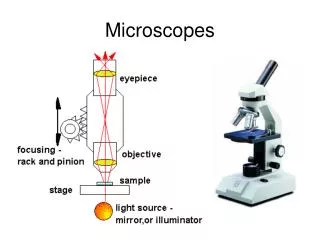
Types of Microscopes • 1. Compound Light Microscope • 1st type of microscope, most widely used • light passes through 2 lenses • Can magnify up to 2000x
Ocular lens Objective lenses
Types of Microscopes • 2. Electron Microscope • Used to observe VERY small objects: viruses, DNA, parts of cells • Uses beams of electrons rather than light • Much more powerful
Types of Microscopes • Transmission Electron Microscope (TEM) • Can magnify up to 250,000x
Types of Microscopes • Scanning Electron Microscope (SEM) • Can magnify up to 100,000x