
Asthma in Children: Presentation and Diagnosis
E N D
Presentation Transcript
ASTHMA PATIENT PRESENTATION AND WORK-UP PATHOPHYSIOLOGY Gail Feinberg, DO, FACOFP, dist. andLiisa A. Russell, MD
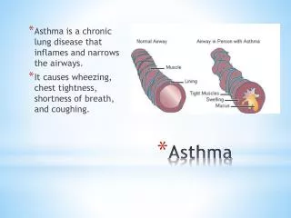
ASTHMA DEFINITION (Harrison): Asthma is a syndrome characterized by airflow obstruction that varies markedly, both spontaneously and with treatment. Asthmatics harbor a special type of inflammation in the airways that makes them more responsive than nonasthmatics to a wide range of triggers, leading to excessive narrowing with consequent reduced airflow and symptomatic wheezing and dyspnea. Narrowing of the airways is usually reversible, but in some patients with chronic asthma there may be an element of irreversible airflow obstruction. DEFINITION (Robbins): Asthma is a chronic disorder of the conducting airways, usually caused by an immunological reaction, which is marked by episodic bronchoconstriction due to increased airway sensitivity to a variety of stimuli; inflammation of the bronchial walls; and increased mucus secretion .
Several Overlapping Classifications: • Atopic • Non-atopic • Intrinsic • Extrinsic • Steroid-dependent • Steroid-resistant • Seasonal • Exercise-induced • Drug-induced • Occupational • Asthmatic bronchitis
6 yr old male with cough • 6 yr old male with 2 months of persistent cough • Mom states it started with a “cold” which resolved, but cough still persisted • Cough is worse at night and with exercise. Mom has tried some OTC cough medicines, none have helped • He never had a cough like this in the past • He had eczema as a child. Family history of allergic rhinitis • No meds currently, no allergies • No recent travel, they have a dog at home, no smokers in the home
ROS • No fevers, chills, weight loss • No rhinorrhea, nasal congestion, sneezing, eye drainage, pharyngitis • Has non-productive cough, no chest pain or SOB, no hemoptysis • No tachycardia • No abd pain, N/V/D • No dry skin or rashes.
DDx? • Allergic rhinitis • Sinusitis • Viral URI • Pneumonia • Bronchiectasis • Tuberculosis • Asthma • Cystic Fibrosis • Foreign body aspiration • Alpha-1 antitrysin deficiency • CHF • Glottic dysfunction • Tumor obstruction
PE • VS: T 98.6, P 96, RR 28, BP 95/66, O2 96% • Gen: WDWN, NAD • HEENT: NCAT, no conjunctival injection, nasal mucosa pink and moist, no polyps, oral mucosa pink and moist, no pharyngeal erythema, no tonsillar enlargement • Neck: supple, no adenopathy • CV: RRR, no murmur, +2 peripheral pulses • Resp: breathing comfortably on RA, no retractions, good air entry, intermittent expiratory wheezes, no crackles or rhonchi • Abd: soft, non-tender, no masses • Ext: no clubbing or cyanosis • Neuro: alert, appropriate for age, normal tone and gait • Skin: no rashes present
What is asthma (simply put)? • A chronic inflammatory disease of the airways • More specifically…. • A disease of the airways characterized by inflammation, hyper-responsiveness, and mucous production that leads to obstruction and air trapping
Asthma Prevalence • One of the most common chronic diseases worldwide • 10-12% of adults, 15% of children • Increasing prevalence in developing countries • Hygiene hypothesis (see slide comments) • Air pollution • Prematurity • Increased awareness?
What causes asthma? • Atopy is the major risk factor • Genetically determined • Triad of atopic dermatitis, allergies and asthma • Triggers of asthma • Allergens- dust mites, pet dander, cockroaches, pollen/grass • Viral upper respiratory infections • Irritants- smoke exposure, perfume, paint fumes/chemicals • Medications- NSAID’s, beta-blockers • Exercise
Asthma: Pathogenesis Extrinsic (allergic), early onset, most common Intrinsic (later in life)
Pathophysiology (intrinsic) • Early phase (within minutes): • Mast cell degranulation stimulated by allergens • Constriction of airway smooth muscle (bronchial hyperrsponsiveness) • Late phase (hours later): • Influx of inflammatory cells (eosinophils, basophils, neutrophils, T lymphocytes) • Increased mucous production
Asthma: Pathogenesis (extrinsic) Note: Asthmatic patient is primed to develop an immune reaction (elevated IgE and Eosinophils are common)
TRIGGERING OF ASTHMA A fundamental abnormality in asthma is an exaggerated T H 2 response to normally harmless environmental antigens.
OVERLAP BETWEEN TYPES OF LUNG DISEASES WITH AIRFLOW OBSTRUCTION
ASTHMA, BAL ASTHMA, BAL MASSIVE EOSINOPHILIA MANY MACROPHAGES CHARCOT- LEYDEN CRYSTAL FROM EOSINOPHIL BASIC PROTEIN GIEMSA PAPANICOLAOU
CURSCHMANN SPIRALS CASTS OF MUCUS GLANDS/DUCTS PAPANICOLAOU-STAIN PAS STAIN
MUCUS CAST OF BRONCHI ASTHMA BRONCHI ARE FILLED WITH TENACIOUS MUCUS
ASTHMA • MORPHOLOGY • Thick basement membrane • Increased airway smooth • muscle • Genetic predisposition to airway remodeling may be present: • ADAM-33 gene polymorphism • (codes a metalloproteinase) • accelerates proliferation of • smooth muscle and fibroblasts • Inflammatory infiltrates and edema in bronchial wall with many eosinophils and mast cells in the cellular phase of an asthma attack • Enlarged mucus glands • Mucus plugs in airways • Charcot-Leyden crystals in • mucus GOBLET CELL HYPERPLASIA THICK BASEMENT MEMBRANE HYPERTROPHIC SMOOTH MUSCLE BUNDLES
ACUTE ASTHMA ATTACK THICK MUCUS MANY EOSINOPHILS IN THE BRONCHIAL WALL THICK BASEMENT MEMBRANE HYPEREMIA HE ALCIAN BLUE-PAS
ASTHMA – KEY PATHOPHYSIOLOGIC CONCEPTS RECAP • Asthma is characterized by reversible bronchoconstriction caused by airway hyperresponsiveness to a variety of stimuli. • Atopic asthma is caused by a T H 2 and IgE-mediated immunologic reaction to environmental allergens and is characterized by acute-phase (immediate) and late-phase reactions. The T H 2 cytokines IL-4, IL-5, and IL13 are important mediators. IL17 and IL9 are also being shown to be important in some asthmatics. • Triggers for nonatopic asthma are less clear but include viral infections and inhaled air pollutants, which can also trigger atopic asthma. • Eosinophils are key inflammatory cells found in almost all subtypes of asthma; other inflammatory cells include mast cells, neutrophils and T lymphocytes. • Airway remodeling adds an irreversible component to the obstructive disease: • - Increased vascularity • - Sub-basement membrane fibrosis (deposition of type I and III collagen) • - Hypertrophy of bronchial submucosal glands • - Increase in the number of goblet cells in the respiratory epithelium • - Bronchial smooth muscle hyperplasia
Clinical Presentation https://www.youtube.com/watch?v=ktmn8QqBHR4 (spirometry) https://www.youtube.com/watch?v=PzfLDi-sL3w (overview)
Clinical Presentation • Cough, dyspnea, wheezing, chest tightness • May present with just a chronic cough • Symptoms are usually worse at night • Often occurs after exposure to a trigger • What is an asthma attack (asthma exacerbation)? • Acute or subacute episodes of progressively worsening symptoms as above
History Questions • What are your symptoms? • How often and when do they occur? How long do they last? • Do symptoms worsen at night, with exercise, or with infections? What are the triggers? • Have you ever been in the ED for your symptoms? Hospital admission? Intubation? • Have you ever used an inhaler (bronchodilator) to help you breathe? • Family history of: asthma, allergies, eczema, nasal polyps?
Physical Exam • Vitals: Tachypnea, Tachycardia, Hypoxia • General: Cyanosis, signs of respiratory distress • Grunting in infants • Head bobbing in infants • Difficulty talking • Tripod position • Nasal flaring • Lip pursing • Accessory muscle use • Retractions • https://www.youtube.com/watch?v=Fmt6JB-W_M8 (Pediatric presentation)
Physical Exam • HEENT: nasal mucosa, nasal discharge, post-nasal drip • Neck: lymphadenopathy, trachea placement • CV: tachycardia, perfusion • Neuro: mental status • Osteopathic: TART findings in T1-T4
Physical Exam • Lungs • Monitor air entry (may have decreased airflow) • Prolonged expiratory phase • Diffuse wheezing • Usually expiratory, but can have both inspiratory/expiratory • Diminished or absence of wheezing in impending respiratory failure • No airflow!!
Asthma Symptom Progression • Episodic (mild)- can be managed in outpatient setting • Chest tightness, non-productive cough • Harsh respirations, mild wheezing, no hypoxia • Severe • Signs of respiratory distress • Wheezing, prolonged respiratory phase • Pulsusparadoxus- fall in SBP >10mmHg during inspiration • Impending respiratory failure • Unable to speak, gasping, cyanosis/hypoxia • Decreased or absence of wheezing
Diagnosis • History and Physical are key! • Confirmation with spirometry (> 5 years old) • Determines if there is airway obstruction • Test reversibility with bronchodilator
Spirometry FVC- normal FEV1 – low FEV1/FVC - low Obstructive pattern Make sure the curves are reproducible Test reversibility
Other diagnostic tests • CXR • Not diagnostic, may show hyperinflation • Can be helpful to rule out pneumonia, other lung pathology • Arterial blood gas (ABG) • May show respiratory acidosis in severe cases • High PCO2, low PO2
Patient example… • CC: A 46-year-old homemaker with a history of allergy to several pollens, animal dander, and house dust is brought to the emergency room in acute respiratory distress. She has several prior admissions because of episodes of severe SOB, usually associated with respiratory infection. • PE: The patient is anxious and has cyanotic lips. T= 37.9°C, P=100, thready, BP= 100/70, R= 22. Respiratory sounds are barely audible. Chest CT, and pulmonary function- and blood gas studies from a previous visit are available. • What are the key clinical features? • What is your clinical impression?
ASTHMA PATIENT HRCT What are the key radiologic features? INSPIRATION: Normal study EXPIRATION: A mosaic pattern of lung attenuation in a patient with asthma. lucent areas (arrows) represent areas of air trapping. Note the increased bronchial markings
SPIROMETRY What is the pattern? The dotted line shows the spirometry after bronchodilator was given. What happened?
PULMONARY FUNCTION STUDIES AND BLOOD GASES DURING A PREVIOUS VISIT What are the key features?
ASTHMA PATIENT IN TROUBLE KEY FEATURES: Hyperinflated lungs and pneumomediastinum. Increased bronchial markings. Hyperinflared alveoli have ruptured and let air escape into the mediastinum.
Hospital course… • In the ER the patient was hypoxemic. The EKG showed right axis deviation. The chest film showed hyperinflated lungs and pneumomediastinum. • The patient was intubated and received treatment directed toward controlling bronchoconstriction and inflammation and restoring fluid and electrolyte balance. • In spite of vigorous treatment the patient expired. The lung tissue was obtained at autopsy. • What are the key morphologic features? • Is the process in the lungs acute or chronic? • Is the process in the lungs reversible?
WHAT HAPPENED TO THE BRONCHIAL EPITHELIUM? WHAT ARE THE CELLS IN THE BRONCHIAL WALL AND IN THE BRONCHIAL LUMEN ?
WHAT HAPPENED TO THE ALVEOLI AND THE ALVEOLAR CAPILLARIES? 1 1 2 2 3 1 3
The patient died More questions: What was the most likely cause of death? Why did this patient die (i.e. what was the mechanism of death)? Was this death preventable?