GIT Bleeding: VGIB LGIB
GIT Bleeding: VGIB LGIB. Dr. Mohamed Shekhani CABM-FRCP. Variceal bleeding:. 1. Clinical decompensation (i.e., ascites, encephalopathy,a previous episode of hemorrhage, or jaundice). Common lethal complication of cirrhosis(50% at diagnosis, 7%/year), particularly with:. Esophageal varices.
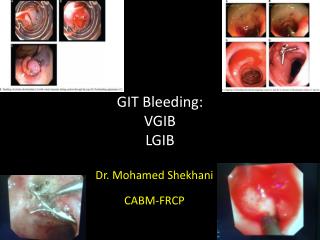

GIT Bleeding: VGIB LGIB
E N D
Presentation Transcript
GIT Bleeding:VGIBLGIB Dr. Mohamed Shekhani CABM-FRCP
Variceal bleeding: 1 Clinical decompensation (i.e., ascites, encephalopathy,a previous episode of hemorrhage, or jaundice). Commonlethal complication of cirrhosis(50% at diagnosis, 7%/year), particularly with: Esophageal varices Gastric fundal varices Or GEV Types Portal hypertensive gastropathy Portal hypertensive Biliopathy
Variceal bleeding: 1 Clinical decompensation (i.e., ascites, encephalopathy,a previous episode of hemorrhage, or jaundice). Commonlethal complication of cirrhosis(50% at diagnosis, 7%/year), particularly with: Treatment of the acute bleeding episode: Mortality 15-20% Primary prophylaxis to prevent a first episode of VH. MANAGEMENT Secondary prophylaxis (prevention of recurrent VH). 60%/year.
VARICES INCREASE IN DIAMETER PROGRESSIVELY Varices Increase in Diameter Progressively No varices Small varices Large varices 7-8%/year 7-8%/year Merli et al. J Hepatol 2003;38:266
PROGNOSTIC INDICATORS OF FIRST VARICEAL HEMORRHAGE Varix with red signs Variceal hemorrhage • Predictors of hemorrhage: • Variceal size • Red signs • Child B/C NIEC. N Engl J Med 1988; 319:983
Portal HT Risk stratification 2 Decompensated liver cirrhosis: Child-Pough or MELD class ( Model of end stage liver disease) Varices or colaterlas detected on imaging studies as Abd U/S,EUS,Dopler Portal HT Risk stratification Gastroesophageal varices. Varices on VCE Plateletes/spleen maximal bipolar diameter<909 Fibroscan measuring liver stiffness predicts portal HT HVPG: gold standard&Best predictor of PHT & EV, but invasive ¬ widely available. >5 mm Hg PHT >10 mm Hg clinically significant
Primary prophylaxis of bleeding eso varices: 3 Propranolol 20mgm*2 untill PR 55/min Indefinite Propranolol Or EBL Sessions every 4 weeks Nadolol 40mgm once daily Untill PR 55/min Indefinite PP of EV bleed Endoscopic band ligation Evey 4 weeks untill total obliteration Follow up: 3 /12 for 1 year, yearly Nadolol 3 MONTHLY For 1 year Then Yearly Indefintely. FU OGD after Obliteration:
Management of acute variceal bleeding: 4 Vasoconstrictor Octreotite Somatostatin Telipresin 5 days Antibiotics: Ceftriaxone Ciprofloxacin 5 days Endoscopic Intervention EBL Sclerotherapy Acute variceal Bleeding. Cyanoacrylate Injection Sclerotherapy For gastric Varices. Sigestaken Tube temponade Esophageal stenting
Secondary prophylaxis( prevention of recurrent) of bleeding EV: 5 Propranolol Same as for primary prophylaxis. Isosorbide propranolo Nadolol Nadolol Same as for primary prophylaxis. Isosorbide dinitrate 10 mgm*10-20 mgm*2 Secondary prophylaxis EBL Same as for primary prophylaxis. EBL for EV Cyanoacrylate for GV Interventional Radiology for GV Cyanoacrylate injection sclerotherapy or IR for gastric varices not EBL.
Portal Hypertensive Gastropathy 14 Definition: • PHT- related ectatic gastric mucosal vessels mostly in fundus • & body of the stomach. Predictors of its presence GEV , Child class& prior variceal endoscopic therapy Prsentation Chronic blood loss leading to IDA rather than acute bleeding Treatment: Iron supplementation;BB,Shunt therapy(surgeryorTIPS) Prophylaxis Same.
Acute Lower Gastrointestinal Bleeding • Bleeding distal to the ligament of Treitz for <less than 3 days. • The colon is the most common site of bleeding. • The incidence increases with age, with mean of 63-77 years. • LGIB accounts for 20% of all episodes of GIB. • Most episodes of LGIB will stop without intervention. • The most common causes of acute LGIB are diverticulosis, angiectasia, ischemic colitis, perianal disease. • The most frequent causes of chronic LGIB are neoplasms, angiectasia, IBD.
Causes in Our locality: Perianal diseases(piles/Fissure) IBD(UC>CD) Infectious colitis Neoplasms(adenoma or cancer) Solitary rectal ulcer syndrome (SRUS) Meckel’sdiverticulum. Ischemic colitis. Angiodysplasia
Hemorrhoids/ fissures: Piles Fissure Bleeding after/or with defecation Pain & bleeding with defecation 1 Careful perianal exam+ anoscopy assist in the diagnosis 2
Initial evaluation/ resuscitation Triage to OP vs Ward vs ICU Acute LGIB: Management algorythm Mild scanty bleeding Anorectal pathology susspected Rigid Anoscopy or sigmoidoscopy to confirm diagnosis Outpatient management Anorectal pathology(piles/fissure) is the most common pathology in our locality But this should be diagnosed on solid basis not to miss serious pathologies as IBD or cancer.
Acute LGIB: Management algorythm Severe bleeding Severe exanguinating bleeding Emergency angiography for bleeding control by gel form or coils Or emergency surgical consult. If emergency angio succeeded just observe for recurrence but if fails refer to surgery SURGERY Severe exanguinating bleeding needs urgent action either emergency surgery or emegency therapeutic interventional radiology.
Moderate severe bleeding Acute LGIB: Management algorythm Consider NGT aspirate Bloody NGT aspirate Risk for UGIB OGD If +ve treat accordingly Most of the cases of LGIB fall in this category & require 1st NGT aspiration & if +ve bloody aspirate , urgent upper GIT endoscopy.
Moderate severe bleeding Acute LGIB: Management algorythm NGT not done or –ve aspirate Polyethelene glycol(PEG) solution laxative for preparation for emergency colonoscopy in few hours. Colonoscopy within 12-24 hours Manage according to colonoscopic findings If the NGT aspirate is not bloody or NGT was not inserted, urgent prep with PEG is needed for urgent colonoscopy within12-24 hours.
Moderate severe bleeding Acute LGIB: Management algorythm On colonoscopy bleeding site & cause is identified so treat as appropriate. If the colonoscopy identifies the site/cause of bleeding the problem is solved
Moderate severe bleeding Acute LGIB: Management algorythm If On colonoscopy there is visual impairment because of ongoing bleeding Angiography. If on colonoscopy there was visual impairment due to bloody field urgent angiography is indicated fordiagnosis & therapy.
Moderate severe bleeding Acute LGIB: Management algorythm On colonoscopy bleeding site not identified but bleeding had stopped OGD Or Repeat colonoscopy Or SI evaluation /Or Others( RBC scan,angiography) for rebleeding. If on colonoscopy the bleeding had stopped & no lesion was identified, upper GI endoscopy is considered(if had already been done) or RBC scan/angigraphy Is done fordiagnosis/treatment specially if bleeding recurred.