Download
1 / 46
510 likes | 805 Vues
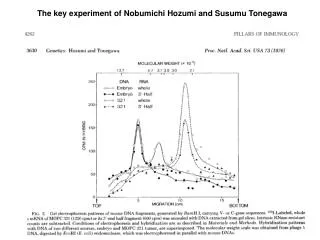
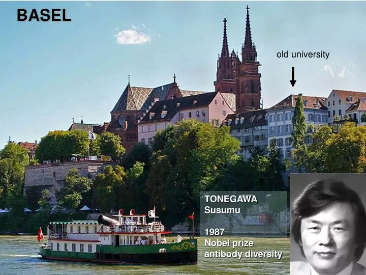
BASEL. old university. TONEGAWA Susumu 1987 Nobel prize antibody diversity. FREIBURG. TAWARA, Sunao 1906 cardiac conducting system. LEISSIGEN / Interlaken Switzerland. EIGER north face - „top of Europe“. IMAI Michiko et al. 23.7.1969 Japanese direttissima route.

E N D
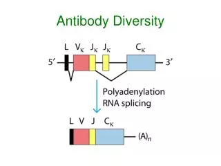
BASEL old university TONEGAWA Susumu 1987 Nobel prize antibody diversity
FREIBURG TAWARA, Sunao 1906 cardiac conducting system
LEISSIGEN / Interlaken Switzerland
EIGER north face - „top of Europe“ IMAI Michiko et al. 23.7.1969 Japanese direttissima route
Teaching Modules in Pathology Education Prof. Dr. Urs-N. Riede, Department of Pathology, University of Freiburg
Textbook: Georg Thieme, Stuttgart – New York first edition 1986 second edition 1988
Textbook: Georg Thieme, Stuttgart – New York third edition 1992 forth edition 1998
Textbook: Georg Thieme, Stuttgart – New York fifth edition 2004 chinese editions 1998, 2004
Color atlas: Georg Thieme, Stuttgart – New York german edition 1998
Color atlas: Georg Thieme, Stuttgart – New York english edition 2008 greek edition 2009
teaching in pathology:aims • The pathologic diagnosis of a disease results from the superposition of formal reaction patterns. • The diagnosis of formal reaction patterns requires the analysis of a few structural and color changes. • The stepwise analysis of the formal reaction patterns elucidates the progress of a disease.
Textbook: Springer Heidelberg – New York – Tokyo 2009BASICS IN GENERAL AND SPECIAL PATHOLOGY • new: • learning modules • structural pattern • contour pattern • consistency pattern • color pattern …
teaching in pathology:modules • Substructuring of a lesion into formal reaction patterns • Pathobiologic explanation of the reaction patterns • Assembling of distinct reaction patterns to a diagnosis • Reconstruction of the pathogenetic sequence • Training by cases of a virtual autopsy
quick-fix diagnosis: facial skin lesions The patient suffered from lupus erythematodes But why did she die?
color red pattern - inflammation - bleeding disorder distribution multiple pattern - septic purpura diagnosis by pattern analysis formal macrofocal pattern - lupus erythema microfocal - purpura
microfocal lung cancer miliary TBC patterns of expansion macrofocal skin erythema
macrofocal secondary tumor patterns of distribution (uni-) macrofocal multifocal microfocal systemic generalised primary tumor inflammation bleeding disorder destruction
spreading patterns lymphatic haematogenous cavitary coagulopathy inflammation inflammation inflammation cancer cancer cancer cancer cancer cancer dis-/continuous inflammation cancer discontinuous cancer
abscess sharp contours lipoma cyst destruction: - ischemia > infarction - inflammation > abscess proliferation: > benignoma retention:> (pseudo) cyst contour patterns fuzzy contours skin erythema melanoma instillation / infiltration: - gaz > emphysema - water > edema - cells > inflammation > malignoma
netlike reticular striped whirled muscle lung lymphangiosis fat interposition fibroma structural patterns diffuse diffus skin erythema
pseudo-membrane ulcer erosion epidermis mucosa subcutis submucosa surface patterns polyp
sphere • large: nodular • small: granular hollow sphere diffuse cystic pseudocystic granular nodular diffuse steric patterns
firm-elastic liquid brittle fluid liquefaction proteolysis soft pulpy soft tissue proteolysis consistency patterns hard fibrosis very hard ossification
color patterns: e.g. liver normal (perfusion) - hemoglobin fatty degeneration icterus bilirubin normal + oxy- hemoglobin leucemia (CLL) cholestasis bile cell infiltration neutral fat cyanosis melanoma siderosis desoxy- hemoglobin melanin hemosiderin
color patterns: white Tyndall-effect because of - coagulation - increase of collagen coagulation myofibroma pleuritis - increase of keratin - increase of fibrin - increase of cells leukoplakia carcinoma
color patterns: red oxyhemoglobin red desoxyhemoglobin red solar erythema livores cyanosis tyrosinogenous yellow-red porphyria / urin tyrosinogenous yellow-red inflammation (chemosis) pheomelanin
- retention - dilatation - inflammation - hyperplasia pre- stenosis normal post- - dysfunction - atrophy patterns of hollow organspatterns of stenosis complications - thrombus/embolus - neoplastic tumor - inflammatory tumor - foreign body - ligatur / accretion direction of removal
case history • 48 year-old patient • cigarette smoker (27 pack years) • onset: flu-like symptoms (caugh, fatigue) • x-ray: suspicion of pneumonia > antibiotics • recurrent pericardial + pleural effusions • back pain • accelerating respiratory insufficiency • cachexia • exitus
- white > cell increase = tumor tumor cut face right lung color pattern of the lesion ? - white > collagen increase - white > fibrin increase - white > keratin increase
- fuzzy > infiltration fuzzy contour infiltration: - water - cells cut face right lung contour pattern of the lesion? - sharp > encapsulation - sharp > destruction - fuzzy > edema
pulmonary lesion color contour consistency surroundings false false false false white fuzzy hard infiltrating lung cancer
cut face left lung formal pattern of the lesion - unifocal pattern - cystic pattern - diffuse pattern - reticular pattern
tumor cut face left lung color pattern of the lesion ? - white > collagen increase - white > fibrin increase - white > necrotic coagulation - white > tumor
progress of the lung cancer form color distribution surroundings false false false false reticular white diffuse infiltrating intrapulmonary tumor spreading (lymphangiosis)
- multinodular pattern surface of the visceral pleura formal pattern of the lesion ? - cystic pattern - reticular pattern - diffuse pattern
tumor surface of the visceral pleura color pattern of the lesion - white > collagen increase - white > fibrin increase - white > necrotic coagulation - white > tumor
- cavitary metastasis surface of the visceral pleura spreading pattern of the lesion - lymphogenous metastasis - hematogenous metastasis - ductogenous metastasis
systemic complications of the lung cancer form color steric spreading false false false false reticular white multinodular cavitary cavitary metastasis
- dark-red > cyanosis cyanosis cut face right lung color pattern of the lesion - bright red > bleeding - wine red > porphyria - yellow red > pheomelanin
stenotic pattern complications of the lesion - poststenotic atrophy - poststenotic dysfunction - prestenotic hyperplasia post- stenotic stenosis prestenotic cut face right lung - prestenotic retention
local complications of the lung cancer color form consistency stenotic pattern false false false false cyanosis diffuse proteolysis prestenotic prestenotic retention pneumonia
pathogenetic sequence of the lesions 1 > 2 > 3 > 4 3 > 2 > 1 > 4 4 > 1 > 3 > 2 1 2 3 4
false evaluation of student back to theory pathogenetic sequence of the lesions 1 > 2 > 3 > 4 3 > 2 > 1 > 4 4 > 1 > 3 > 2 2 > 4 > 1 > 3 2 4 1 3
tissue lesion teacher: formalpathogenetic patterns false student: explorative partialdiagnoses pathologic fulldiagnosis student‘s evaluation number of „false“
primary lesion expansion: macrofocal > primary lesion color: white > tumor contour: fuzzy > malignoma stenosis:prestenotic > secondary lesion > mucus retention color: cyanosis > no oxygenation consistency: pulpy > inflammatory proteolysis secondary lesion