Download
1 / 25
250 likes | 276 Vues
Explore the characteristics, classifications, and occurrences of epithelial tissues in the body, including simple squamous, cuboidal, columnar, and transitional types. Learn about their primary functions and cellular components.

E N D
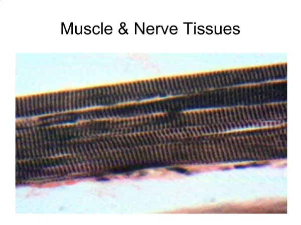
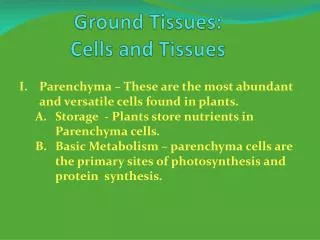
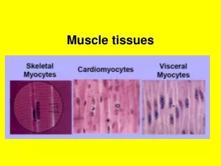
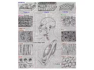
THE GROUND TISSUES EPITHELIAL TISSUES CONNECTIVE AND SUPPORTIVE TISSUES MUSCLE TISSUES NERVOUS TISSUE
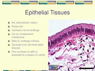
HISTOLOGY 1.3.: EPITHELIAL TISSUES 1. SURFACE EPITHELIA • Diverse group of tissues, cover or line external and internal body surfaces, • cavities and tubes, thus, function as interfaces between different • biological compartments. • Epithelia mediate a wide range of activities: • selective diffusion • absorption • secretion • physical protection • sensory function • Origin of the epithelial tissues: • ectoderm (corneal epithelium, epidermis of skin, glands) • entoderm (alimentary tract, intestinal glands, liver, pancreas) • mesoderm (kidney, male and female reproductive system, • endothelium, mesothelium)
Blood supply of the epithelial tissue: • normally it is avascular • exception: pharyngeal epithelium of frog • urinary bladder of rabbit • Nerve supply of the epithelial tissue: • it is an innervated tissue containing free nerve endings • and nerve endings associated to specialized cells • of sensory function • Tissue components of epithelial tissues: • Cells (squamous, cuboidal, columnar) closely packed • Intercellular matrix is reduced to the glycocalyx of the adjacent cells
Membrane modifications of the apical surface of epithelial cells: cilia Membrane specialization of the basal membrane surface of epithelial cells: basal striation + basal lamina (green arrows)
CLASSIFICATION OF EPITHELIAL TISSUES FUNCTIONAL BASIS: SURFACE EPITHELIA GLANDULAR EPITHELIA ABSORPTIVE EPITHELIA PIGMENT EPITHELIA SENSORY EPITHELIA
SURFACE EPITHELIA: Further classification: on the basis of the number of cell layers on the basis of the shape of the cells Simple epithelia: a single layer of cells simple squamous simple cuboidal simple columnar Pseudostratified epithelia: single layer of cells, with nuclei in more than one layer pseudostratified columnar transitional Stratified epithelia: several layers of cells on top of each other stratified squamous (non-keratinized, keratinized) stratified cuboidal stratified columnar
OCCURRENCE OF SIMPLE SQUAMOUS EPITHELIUM IN THE BODY • PULMONARY ALVEOLI • PARIETAL LAYER OF BOWMAN’S CAPSULE – KIDNEY • THIN SEGMENTS OF HENLE’S LOOP – KIDNEY • RETE TESTIS • INNER ASPECT OF THE TYMPANIC MEMBRANE IN THE MIDDLE EAR • ENDOTHELIUM LINING THE BLOOD AND LYMPHATIC VESSELS • SEROUS MEMBRANES LINING BODY CAVITIES AND INTERNAL ORGANS
SOME EXAMPLES: Cross-section of the simple squamous epithelium: Kidney glomerulus, Bowman’s capsule Mesothelial lining of the peritoneal cavity: „fried-egg” appearance
Endothelium: lining of the blood vessels and heart (arrows). Note the flattened nuclei The alveoli of the lung are composed of similar cells
OCCURRENCE OF SIMPLE CUBOIDAL EPITHELIUM IN THE BODY • LINING THE FOLLICLES IN THE THYROID GLAND • THE SURFACE OF OVARY • CHOROID PLEXUS – BRAIN • THE CAPSULE OF LENS – EYE • PIGMENTED EPITHELIUM OF RETINA – EYE • DUCTS OF MANY GLANDS • SECRETORY ACINI OF MANY GLANDS • SEVERAL SEGMENTS OF KIDNEY TUBULES
Kidney: proximal and distal convoluted tubules, collecting tubules are lined by simple cuboidal epithelium Proximal tubules in cross-section and in longitudinal section (arrow)
The follicles of the thyroid gland are formed by simple cuboidal cells
OCCURRENCE OF SIMPLE COLUMNAR EPITHELIUM IN THE BODY • DIGESTIVE TRACT FROM THE CARDIA OF THE STOMACH TO THE ANUS • LARGER EXCRETORY DUCTS OF SOME GLANDS • UTERUS (CILIATED) IN HUMAN • OVIDUCT (CILIATED) IN HUMAN • PULMONARY BRONCHI (CILIATED) - LUNG • PARANASAL SINUSES – NOSE • CENTRAL CANAL OF SPINAL CORD (EPENDYMA)
Pseudostratified epithelia: pseudostratified columnar epithelium
OCCURRENCE OF PSEUDOSTRATIFIED COLUMNAR EPITHELIUM • THE MALE URETHRA • EPIDIDYMIS – MALE GENITAL ORGAN • TRACHEA (CILIATED) • PRIMARY BRONCHI (CILIATED) – LUNG • THE AUDITORY TUBE • PART OF THE TYMPANIC CAVITY – EAR • LACRIMAL SAC
Urinary bladder, fixed in empty state: note the thickness of the urothelium: 8-10 cell layers (arrow) Urinary bladder, fixed in full state: note the thickness of the urothelium: 4-5 cell layers (arrow)
The luminal surface of the umbrella cells is covered by a specialized cell membrane called calyx, or crusta
Stratified epithelia: stratified squamous non-keratinized epithelium • Layers from the basal lamina to the surface: • stratum basale/germinativum: columnar cell layer with mitotic division • stratum spinosum: cuboidal poligonal cell layers with many desmosomes • stratum planocellulare: fusiform or flattened cell layers • Occurrence: cornea, internal body surfaces, e.g. esophagus (see above)
Stratified epithelia: stratified squamous keratinized epithelium 4. 1. 2. 3. 5. Str. corneum Layers from the basal lamina to the surface: 1. stratum germinativum, 2. stratum spinosum, 3. stratum granulosum (fusiform cells with keratohyalin granules), 4. stratum lucidum (flattened layer with eleidin), 5. stratum corneum (dead cell layer) Occurrence: epidermis of skin, beginning and end-portions of the gastrointestinal tract
STRATIFIED CUBOIDAL EPITHELIUM VERY RARE: TWO ROWS OF CUBOIDAL CELLS ON TOP OF EACH OTHER Duct of the sweat gland (cisterna lactis of teat- not shown) STRATIFIED COLUMNAR EPITHELIUM: Cavernous urethra, Large excretory ducts of some glands, Larynx (ciliated), Fetal esophagus (transiently)