Understanding Protein Digestion by Pepsin: A Laboratory Simulation Study
This laboratory simulation explores the digestion of proteins by the enzyme pepsin, produced in the stomach. We examine the role of hydrochloric acid (HCl) in activating pepsin from its inactive form, pepsinogen, and its effectiveness at various pH levels and temperatures. BAPNA, a synthetic protein substrate, demonstrates pepsin's activity through color change. Results show the impact of incubation time and temperature on enzyme activity, highlighting the vital conditions for optimal protein digestion in the stomach.

Understanding Protein Digestion by Pepsin: A Laboratory Simulation Study
E N D
Presentation Transcript
GI Laboratory Simulation: Digestion Group 1: Aguila, Alog, Alejandro, Almajar, Angeles, Araño, Balictar, Buemio, De los Arcos, Escobillo, Manuel, Peña, Rabanal, Rivera,Ronquillo, Siazon,Sutingco,Tagalog, Tamayo, C. Uy, Yeo, Yu
Activity 3 Protein Digestion by Pepsin
Pepsin • A protein-digesting enzyme • Produced by chief cells of the stomach glands • Hydrolyzes proteins to small fragments • Amino acids and some polypeptides
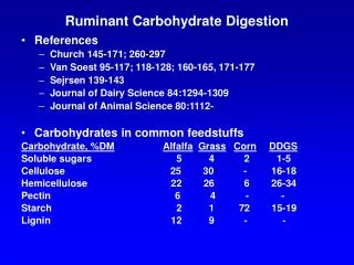
Gastric Secretion: Background • 2 types of glands in the stomach mucosa: • Oxyntic (acid-forming) glands • aka gastric glands • Inside surfaces of the body and fundus • Secrete Hydrochloric acid, pepsinogen, intrinsic factor, mucus, gastrin • Composed of 3 types of cells • Mucous neck cells: secrete mucous • Peptic chief cells: secrete pepsinogen • Parietal (oxyntic) cells: secrete HCl and intrinsic factor • Pyloric glands
Pepsinogen and Pepsin • Pepsinogen: inactive form of Pepsin • Becomes activated into pepsin when it comes into contact with Hydrochloric acid • Pepsin • An active proteolytic enzyme in a highly acidic medium • Optimum pH 1.8 – 3.5 • Above pH of 5: almost no proteolytic activity and may become completely inactivated
Protein Digestion in Stomach • Requires stomach juices to be acidic for it to be active • HCl secreted by parietal (oxyntic) cells in glands • When mixed with stomach contents, pH becomes 2.0-3.0 (highly favorable for pepsin activity)
Protein Digestion by Pepsin • Pepsin can digest collagen • Important for digestion of meat and other meat proteins • Only initiates protein digestion • 20% of total protein digestion • Converts protein to: • Proteases • Peptones • Few polypeptides • Breaks down protein by hydrolysis at peptide linkages between amino acids
Lab Simulation Acitivity 3 Protein Digestion by Pepsin
Procedure • BAPNA: synthetic protein • Transparent and colorless in solution • Will turn yellow if active pepsin (or any protein digesting enzyme) is present • Pepsin will digest BAPNA • Pepsin Incubation • Boil Test tube 1 • Pepsin Assay • Spectrophotometer: to measure optical density
Run 1 Incubation Temperature: 37°C Incubation Time: 60 minutes
Run 2 Incubation Temperature: 37°C Incubation Time: 30 minutes
Run 3 Incubation Temperature: 10°C Incubation Time: 60 minutes
Analysis 1. Which pH provided the highest pepsin activity? How does this correlate to the location of pepsin in the body? 2. Would pepsin be active in the mouth? Explain. • pH 2.0 • Pepsin obtains optimal activity in a highly acidic medium (pH 1.8-3.5). When pH exceeds 5.0, very little or no proteolytic activity will take place. • This pH correlates with the pH in the stomach which has an acidic pH of 2.0-3.0 when HClmixes with stomach contents • No. • The normal pH of the mouth stays close to neutral (7.0). This pH however, can change temporarily when certain foods are digested. The pH of the mouth does not reach the ideal acidity in order for pepsin activity to take place (pH 1.8 – 3.5).
Analysis 3. How did the results of tube 1 compare with those of tube 2? Test tube 1 displayed an optical density of 0.00 while test tube 2 displayed an optical density of 0.40. The solution in test tube 1 remained clear whereas the solution in test tube 2 turned yellow, evidence that BAPNA in test tube 2 was digested. 4. Tubes 1 and 2 contained the same substances. Explain why their optical density measurements were different. The boiling of the solution in test tube 1 gave rise to the difference in optical density measurements between tubes 1 and 2. The boiling of the solution in test tube 1 caused the denaturing of pepsin, rendering this inactive and unable to digest BAPNA.
Analysis • 5. Did the pepsin or deionized water contain any contaminating digested BAPNA? Which tubes confirm this? • No. The pepsin or deionized water did not contain contaminating digested BAPNA. Test tubes 3 and 4 confirm this with 0.00 optical density. • 6. What do you think would happen if you reduced the incubation time to 30 minutes? How did this affect optical density results? • Reducing the incubation time to 30 minutes reduced the optical density due to the reduced amount of time for digestion of BAPNA.
Analysis 7. What do you think would happen if you decreased the temperature to 10°C? What effect would this have on pepsin activity? Why? What effect did boiling have on pepsin? • Decreasing the temperature to 10°C would decrease pepsin activity and would decrease the amount of digested BAPNA. • Decreasing the temperature would, in effect, decrease optical density. • Pepsin works best at body temperature (~37°C) • Boiling led to the denaturation of pepsin rendering this inactive.
Analysis • What is the substrate in the experiment? • The substrate used in the experiment was BAPNA • BAPNA: Nα-benzoyl-DL-arginine-p-nitroaniline • What was the significance of using 37°C for incubation? • 37°C was used for incubation because the experiment tried to simulate the activity of pepsin in the body. 37°C is representative of normal body temperature.
Activity 4 Pancreatic Lipase Digestion of Fats and the Action of Bile
Lipase • Necessary for the absorption and digestion of nutrients in the intestines. • Responsible for breaking down lipids (fats), in particular triglycerides. Once broken down into smaller components, triglycerides are more easily absorbed in the intestines. • Produced in the pancreas but is also produced in the mouth and stomach. Most people produce sufficient amounts of pancreatic lipase.
Lab Simulation Acitivity 4 Pancreatic Lipase Digestion of Fats and the Action of Bile
Analysis 1. Explain the difference in activity between test tubes 1 and 2. 2. Can we determine if fat hydrolysis has occurred in tube 6? Explain. • The presence of bile is needed to emulsify fat into droplets • As fat is digested, it produces organic acid end products that lower pH levels. • Bile salts in test tube 1 more effectively broke down fat and produced organic acid end products, and thus lower the solution's pH more than test tube 2. • Some fat hydrolysis has still occurred • Bile salts and lipase are still present, fat was still hydrolyzed into its component fatty acids. • Minimal change in pH is caused by organic acids produced in the reaction
Analysis 3. Which pH resulted in maximum lipase activity? 4. Can we determine if fat hydrolysis has occured in tube 5? Explain. • pH 7.0 resulted in maximum lipase activity. • This pH correlates with the optimum pH for pancreatic lipase [8.0] • No. • However, studies have shown that below pH 3.0, human pancreatic lipase has irreversibly lost enzyme activity and its ability to bind to lipid emulsions is abolished • Most likely, no reactions occurred
Analysis 5. In theory, would lipase be active in the mouth? In the stomach? 6. What is the substrate, and what subunit is formed in this experiment?. • Mouth - pH 7 • Yes, lipase can be active in the mouth because the pH is near the optimum pH • Stomach – pH 2-3 • No, lipase would be inactive in the stomach as it is well below the optimum pH Vegetable oil (fat) Bile salts Lipase Monoglycerides and fatty acids
The enzyme lipase is located in the small intestine, where the pH is neutral. Analysis 16. Describe the activity of lipase with & without the bile salts. 18. What pH resulted in the maximum pancreatic lipase activity?. • The activity of lipase is enhanced with the addition of bile salts. This can be seen in the greater decrease in pH of tube 1 over that of tube 2. • Bile activity is a physical process. The fat particles are simply being broken down into smaller fat particles, to aid the subsequent activity of lipase. • pH 7.0 • The enzyme lipase is located in the small intestine, where the pH is neutral. 19. How does this optimal pH correlate to the enzyme’s location in the body?. 17. Is the activity of bile a chemical or a physical process?
References • Guyton, Arthur, and John Hall. Textbook of Medical Physiology. 11th ed. Philadelphia, Pennsylvania: Elsevier Saunders, 2006. Print. • "Pepsin." Enzyme Explorer. 2009. Sigma-Aldrich Co., Web. 25 Nov 2009. <http://www.sigmaaldrich.com/life-science/metabolomics/enzyme-explorer/analytical-enzymes/pepsin.html>.
http://www.worthington-biochem.com/introbiochem/effectspH.htmlhttp://www.worthington-biochem.com/introbiochem/effectspH.html • Ranaldi, S et al. 2008. Lid Opening and Unfolding in Human Pancreatic Lipase at Low pH Revealed by Site-Directed Spin Labeling EPR and FTIR Spectroscopy. Biochem 48 (3), pp 630–638