Download
1 / 31
360 likes | 827 Vues
MB207 MOLECULAR CELL BIOLOGY The Cell Cycle and Programmed Cell Death. An overview of the cell cycle. Prophase. Metaphase. Anaphase. Kinetochore microtubules attach to chromosomes. Chromosomes move to midline/metaphase plate. Centrosomes migrate. Spindle fibers form.

E N D
MB207 MOLECULAR CELL BIOLOGY The Cell Cycle and Programmed Cell Death
Prophase Metaphase Anaphase • Kinetochore microtubules attach to chromosomes. • Chromosomes move to midline/metaphase plate. • Centrosomes migrate. • Spindle fibers form. • Chromatin condenses. • Chromosomes separate. • Daughter chromosome • move to poles. Telophase Cytokinesis • Chromosomes reach poles and decondense. • Nuclear envelope reforms. • Spindle fibers disappear. M phase
The cell-cycle control system triggers the major processes of the cell cycle
The significant of cell cycle checkpoints • The DNA replication checkpoint • mitosis is prevented if DNA is not replicated properly. • The spindle attachment checkpoint • Chromosome separation is delayed if the chromosome are not properly attach. • DNA damage checkpoint • Progression through G1 and G2 is slowed if DNA damage is detected • These checkpoints operate through negative intracellular signals • For example, the cell cycle control system receives a ‘stop-anaphase’ signal from each chromosome until the mitotic spindle is attached. When all are attached all of the stop signals are gone and the process is allowed to proceed.
The cell-cycle control system is based on cyclically activated protein kinases • Cdk control cell-cycle system. • A complex of cyclin and Cdk acts as protein kinase to trigger specific cell-cycle events. • Without cyclin, Cdk is inactive.
Structural basis of Cdk activation • In the inactive state, without cyclin bound, the active site is blocked by a region of protein called the T-loop (red). • The binding of cyclin causes the T-loop to move out of the active site, resulting in partial activation of Cdk2. • Phosphorylation of Cdk2 at threonine residue in the T-loop further activates the enzyme by chnaging the shape of T-loop, improving the ability of the enzyme to bind its protein substrates. Cyclin not only activate the Cdk, but direct them to particular substrates
Regulation of Cdk activity by inhibitory phosphorylation • In addition to cyclin activation, other layers of regulation are imparted by phosphorylation/ dephosphorylation Inhibition of cyclin-Cdk complex by CKI • Inhibitor protein binding can also regulate cyclin/ Cdk activity. p27 is a Cdk inhibitor (CKI). CKIs function primarily in the G1-S transition.
The cell-cycle control system depends on cyclical proteolysis • Ubiquitin mediated proteolysis is responsible for cyclin degradation and Cdk inactivation at the major cell cycle transitions. The regulated step in this process is the final addition of ubiquitin to the substrate protein by an ubiquitin protein ligase. A ubiquitin protein ligase complex called SCF is responsible for initiating S-phase by the destruction of G1-S cyclins and certain CKIs. The ubiquitin protein ligase complex called anaphase-promoting complex (APC) is responsible for initiating Anaphase by the destruction of M cyclins and regulators.
Control of proteolysis by anaphase-promoting complex (APC) • Proteolysis is the directed degradation of proteins by cellular enzymes called proteases or by intramolecular digestion.
Intracellular control of cell cycle events • Each cyclin/ Cdk complex serves as a molecular switch to initiate a specific cell cycle event • S-phase cyclin-Cdk complexes (S-Cdks) initiates DNA replication once per cycles
Control of DNA replication initiation • The ORC complex nucleates the association machinery required for replication beginning with Cdc6 (cdc= cell division control). • This complex is ready to replicate DNA but is held in check by Cdc6. • Activation of S-Cdk phosphorylates Cdc6, leading to its degradation and initiation of DNA replication.
Activation of M-Cdk • The activation of M-phase cyclin-Cdk complexes (M-Cdks) triggers entry into mitosis. Activation begins by the accumulation of the M-cyclins (cyclin B) due to a reduction in degradation. The activity of M-Cdk is held in check by a complex set of regulatory factors. The brakes are removed by activity of a phosphatase called Cdc25. • At the end of G2, the cell has a stockpile of the inactive M-Cdk complex. The initial phosphorylation of Cdc25 is though to occur by a protein kinase known as polo kinase. A positive feedback loop causes a near binary switch to ‘on’ at the G2/M transition.
M-Cdk prepares the duplicated chromosomes for separation • Phosphorylation of the condensin complex induces chromosome condensation. • Phosphorylation of the nuclear lamins induce nuclear envelope breakdown. • Phosphorylation also triggers microtubule rearrangement necessary for spindle formation. • Phosphorylation of Golgi stacking factors causes Golgi vesiculation. Exit from mitosis requires the inactivation of M-Cdk • Inactivation of this activity by APC mediated degradation of M-cyclin allows rapid reversal of these processes by constitutive phosphatases.
An overview of the cell-cycle control system p53 = tumor suppressor gene
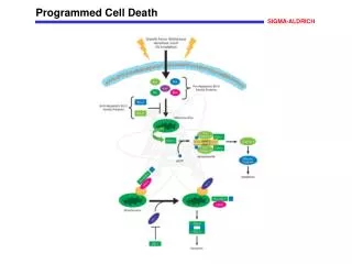
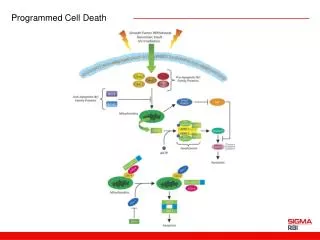
How DNA damage arrests the cell cycle in G1 Mdm2 is an important negative Regulator of the p53 tumor suppressor • When DNA is damaged, protein kinases that phosphorylate p53 are activated. • Mdm2 normally binds to p53 and promotes its ubiquitylation and destruction in proteasomes. • Phosphorylation of p53 blocks its binding to Mdm2. As a result, p53 accumulates to high levels and stimulates transcription of the gene that encodes the CK1 protein p21. • The p21 binds and inactivates G1/S-Cdk and S-Cdk complexes, arresting the cell in G1. What happens if the DNA damage is too severe? The damaged cell commits suicide (apoptosis) in multicellular organisms.
Programmed cell death (apoptosis) - A natural process of self-destruction in certain cells • Apoptosis is a vital process to the overall good of a multicellular organism. Up to half of the neurons in a developing vertebrate organism die soon after they are formed (target selection). Some tissues are formed and most are maintained at a consistent size by selective apoptosis. Sculpting digits by apoptosis Apoptosis during metamorphosis
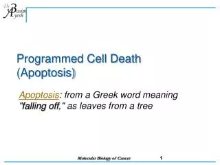
Apoptosis: Programmed Cell Death • Definitions: Apoptosis vs Necrosis • Necrosis derived from Greek “nekrosis”, meaning “deadness”. Necrosis is lethal cell injury or accidental cell death in the living organism. • Apoptosis derived from Greek “apo”, meaning “away from” and “ptosis”, meaning “to droop” or “to fall”. This is programmed cell death
Apoptosis is mediated by an intracellular proteolytic cascade • The primary mechanism of destruction is the activation of proteases called caspases. Most cells have inactive pools of the proteases that are tightly maintained in the off state, mainly in the form of an inactive proenzyme. • The cascade is though to be set in motion by initiator procaspases that are slightly activated by association and aggregation by adaptor proteins. • This activation is triggers by the close proximity or a slight conformation change upon aggregation. Once activated, these initiator procaspases propagate the cascade of activation.
Caspase cascade involved in apoptosis • Each suicide protease is made as an inactive proenzyme (procaspases) • Activated by proteolytic cleavage by another member of the caspase family. • Two of the cleaved fragments associate to form the active site of the caspase. • The active enzyme is thought to be a tetramer of two of these units. • a) Each activated caspase molecule can cleave many proscaspase molecules and activating them. • b) These can activate even more procaspase mlecules. • An initial activation of a small number of procaspase molecules (called initiator caspases) can lead via an amplifying chain reaction (a cascade). • To the explosive activation of a large number of procaspase molecules. • Some of the activated caspases (called effector caspases) then cleave a number of key proteins in the cell, including specific cytosolic proteins and nuclear lamins, leading to the controlled death of the cell.
Induction of apoptosis by extracellular stimuli • A killer lymphocyte carrying the Fas ligand binds and activates Fas proteins on the surface of the target cell. • Adaptor proteins bind to the intracellular region of aggregated Fas proteins, causing the aggregation of procaspase-8-molecules. • The activated caspase-8 molecules then activate downstream procaspases to induce apoptosis. • Then cleave one another to initiate the caspase cascade. • Some stressed or damaged cells kill themselves by producing both the Fas ligand and the Fas protein, thereby triggering an intracellular caspase cascade.
Induction of apoptosis by intracellular stimuli • Mitochondria release cytochrome c that binds to and causes the aggregation of the adaptor protein Apaf-1. • Apaf-1 binds and aggregates procaspase-9 molecules that leads to the cleavage of these molecules and the triggering of a caspase cascade. • Triggering apoptosis requires p53, which can activate the transcription of genes that encode proteins that promote the release of cytochrome c from mitochondria. • Other proteins that contribute to apoptosis are also released from the mitochondrial intermembrane space. These proteins belong to the Bcl-2 family.
Extracellular survival factors suppress apoptosis (Protein kinase B) • IAP seems to bind to apoptosis directly and inhibit their activation.
Growth Factor Receptors • Growth factors regulate growth, proliferation, cellular differentiation and survival. • important for regulating a variety of cellular processes. • act as signaling molecules between cells. Examples are cytokines and hormones that bind to specific receptors on the surface of their target cells. • These are all deregulated in cancer.
Some extracellular signal proteins inhibit cell growth, cell division and survival