Download
1 / 31
310 likes | 484 Vues
Pathophysiology of brain injury. F. Della Corte – C. Maestrone Intensive Care Unit – University of Novara -School of Medicine. Objectives. -To describe which are the common pathophysiological features shared by head injury and stroke -To define the mechanisms of hypoxic-ischaemic

E N D
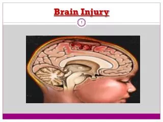
Pathophysiology of brain injury F. Della Corte – C. Maestrone Intensive Care Unit – University of Novara -School of Medicine
Objectives -To describe which are the common pathophysiological features shared by head injury and stroke -To define the mechanisms of hypoxic-ischaemic damage at neuronal level -To stress the importance of ischemia in the determination of severity in the outcome in head injured patients -To define the consequences of ischemic events in the adult
Ischemic stroke vs Head injury Central core Peripheral penumbra In most of the presentations molecular mechanisms are basically the same though operating in: -different sequences -different time courses -different intensities
Deterioration of CBF due to progressive damage of arterial blood supply Activation of cytotoxic processes secondary to formation and/or release of neurotoxic mediators compound and development of tissue acidosis Factors contributing to the increase of irreversibly damaged brain parenchyma
Cellular injury during ischemia • Inadequate Energy supply • Deterioration of Ion Gradients • Consequences of calcium overload
Mild to moderate ischemia Severe ischemia Advanced ischemia Loss of function causes accumulation of glutamate and aspartate which bind to NMDA receptors Insufficient oxygen and glucose Influx of water Na+ Cl- Cytotoxic edema Inadequate energy supply Influx of Ca2+ Influx of water Na+ Ca2+ Irreversible cellular injury Failure of neuronal activity Regional brain dysfunction Anaerobic metabolism Destruction of cell components Formation of free radicals, eicosanoids and leukotrienes Accumulation of lactic acid and H+ compromises neuronal integrity
Cellular injury during ischemia - Inadequate energy supply Ischemia ( O2,glucose) ATP Lactic acid Depolarisation Failed homeostatic mechanisms [H+] [Na+] i [K+] i [Cl-] i VCR [Ca2+] i Neurotransmitters Free Fe2+ LCR Glutamate NA DA Free radicals Lipolysis NO synthesis Auto-oxidation Arachidonic acid Proteolysis Glial injury Free radicals IRREVERSIBLE INJURY
Ischemia and brain injury Prognosis in head injury has been strictly correlated with: -the degree -the duration of the ischemia More than 90% of authopsies on HI pts showed ischemic lesions of different severity Graham D.I., Adams J.H. Ischemic brain damage in fatal head injuries. Lancet 1:265-266, 1971
Vasospasm Intracranial hypertension Arterial hypotension Posttraumatic cerebral ischemia Focal tissue compression from intracranial hematomas Brain edema and swelling
CBF ml/100g/min Time course and CBF in head injury 50 . . . 45 . 40 . . . . . . 35 30 Phase I II III 25 Day 0 1 2 3 4 5 6 7 8 9 10 11 12 13 Martin NA, Patwardhan RV, et al: Characterization of cerebral hemodynamic phases following severe head trauma: hypoperfusion, hyperemia, and vasospasm. J Neurosurg 87: 9-19, 1997
CBF ml/100g/min % ischemia Time course and CBF in head injury . . 40 40 . . . . 30 35 . . . 20 30 10 25 . 20 0 6 12 18 24 30 36 42 48 hours after injury Bouma GJ, Muizelaar JP, Choi SC, et al: Cerebral circulation and metabolism after severe traumatic brain injury: the elusive role of ischemia. J Neurosurg 75: 685-693, 1991
Motorscore = 1,2 = 3,4,5 AJDO2 ml/100ml 9.0 Time course and CBF in head injury . 8.0 . 7.0 . 6.0 . . 5.0 . 4.0 3.0 hours Bouma GJ, Muizelaar JP, Choi SC, et al: Cerebral circulation and metabolism after severe traumatic brain injury: the elusive role of ischemia. J Neurosurg 75: 685-693, 1991
Time course and CBF in head injury % 100 90 CBF (ml/100g/min) 80 70 60 > 55 35 to 55 < 35 50 40 30 20 10 0 I II III Phase Martin NA, Patwardhan RV, et al: Characterization of cerebral hemodynamic phases following severe head trauma: hypoperfusion, hyperemia, and vasospasm. J Neurosurg 87: 9-19, 1997
SEQUENTIAL ACTIVATION OF CEREBROVASCULAR RESPONSES Survival CBF (ml/100g/m) Ischemic threshold Death 50 40 30 20 10 0 1 2 3 4 5 6 7 8 9 10 days post injury Bullock MR et Al J. Neurotrauma 1996; 13; 643-5
SEQUENTIAL ACTIVATION OF INJURY PROCESSES ICP mechanisms Cytotoxic edema Vascular engoargement Vasogenic edema 0 1 2 3 4 5 6 7 8 9 10 days post injury Bullock MR et Al J. Neurotrauma 1996; 13; 643-5
Time course of jugular venous desaturations Gopinath SP: J Neurol,Neurosurg and Psy 1994; 57:717-723 % desaturations (SjO2 < 50% for ten minutes or more)
CBF and incidence of jugular venous desaturations Gopinath SP: J Neurol,Neurosurg and Psy 1994; 57:717-723 ml/100g/min
Oxygen and glucose metabolism after head injury 100 50 0 % Metabolicratio = CMRO2/CMRglu Bergsneider: J Neurosurg 86; 241-251, 1997
Cerebral Blood Flow • 39.9 + 11.2 (Schroeder, 1995) • 42.5 + 15.8 (Mc Laughlin, 1996) • Vasoreactivity 0.4-9.1% 29.3 + 16.4 Mc Laughlin, 1996 Contusion Perilesional edema CT-normal tissue
Brain oxygen tension Van den Brink, Neurosurgery 46; 868-878, 2000
Glutamate Days after injury mM Yamamoto: Acta Neurochir S75: 31-34
Potassium Potassium • Contusion • No contusion mM hours Doppenber EMR: Determinants of cerebral extracellular potassium after severe human head injury. Acta Neurochir 1999; S75: 31-34
Framework of stroke Stroke Infarction 85% Hemorrhage 15% Cerebrovascular disease 80% Intracerebral Cardiogenic embolism 15% Subarachnoid Other unusual 5%
Atherosclerosis and thrombus formation Physiological subtypes of thrombotic-related ischemic stroke Primary large vessel occlusive disease Embolism -arterial atherothrombosis -cardiogenic atrial fibrillation myocardial infarction/ mural thrombus cardiomyopathy prosthetic valves -”paradoxical” (deep vein thrombosis) -atherothrombosis -dissection -arteritis -migraine -drug-induced -etc Thrombosis Primary small vessel occlusive disease -”lacunar” (i.e. microatheroma/lipoyalinosis -arteritis -eclampsia -drug-induced -antiphospholipid antibodies Rotthrock JF In Hemostasis and Thrombosis: Philadelphia, JB Lippincott Company, 1994
Atherosclerosis and thrombus formation Oxydation of LDL cholesterol Monocyte/Macrophage Smooth muscle cells Endothelial cells Free radical release Oxidize LDL cholesterol Scavenger receptor Cytotoxicity Foam cell formation Promote endothelial injury Recruit monocytes Inhibit macrophage egress De Graba TJ in Barnett (eds): Stroke:Pathophysiology, Diagnosis and Management - New York - Churchill Davidson, 1992
Atherosclerosis and thrombus formation Minimal endothelial injury Role of Monocytes and T Lymphocytes in the transformation to foam cells Smooth muscle cell migration and proliferation Platelets adhesion
Atherosclerosis and thrombus formation (2) Plaque fissuring and Formation of platelet thrombus I - Platelets activation II - Platelets adhesion III - Activation of coagulation cascade Thrombus formation
Atherosclerosis and thrombus formation Potential outcome of plaque fissuring 1)fibrotic organization 2)intraintimal and intraluminal thrombosis 3)occlusive thrombosis
Evolution of Cerebral Atherothrombosis The ischemic penumbra
Cerebral Embolism formation I II III Cardiac Sources
Any question from the floor ? • Short !! • Easy to understand!!! • …and to be replied !!!! Please