S2.
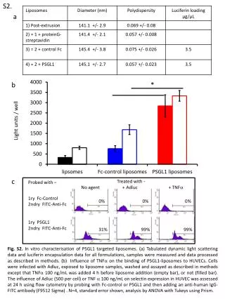
S2. a. b. *. Light units / well. c. Treated with - No agent + Adluc + TNF . Probed with – 1ry Fc -Control 2ndry FITC-Anti- Fc 1ry PSGL1 2ndry FITC-Anti- Fc. 0%. 0%. 0%. 31%. 99%. 99%.

S2.
E N D
Presentation Transcript
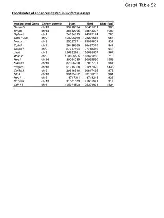
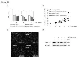
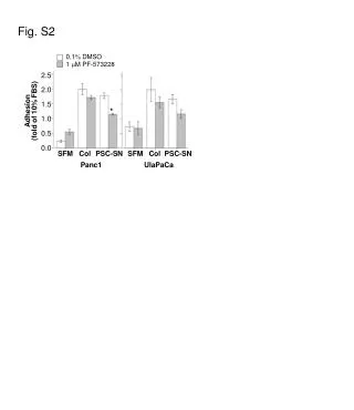
S2. a b * Light units / well c Treated with - No agent + Adluc + TNF Probed with – 1ry Fc-Control 2ndry FITC-Anti-Fc 1ry PSGL1 2ndry FITC-Anti-Fc 0% 0% 0% 31% 99% 99% Fig. S2. In vitro characterisation of PSGL1 targeted liposomes. (a) Tabulated dynamic light scattering data and luciferin encapsulation data for all formulations, samples were measured and data processed as described in methods. (b) Influence of TNF on the binding of PSGL1-liposomes to HUVECs. Cells were infected with Adluc, exposed to liposome samples, washed and assayed as described in methods except that TNF 100 ng/mL was added 4 h before liposome addition (empty bar), or not (filled bar). The influence of Adluc (500 per cell) or TNF 100 ng/mL on selectin expression in HUVEC was assessed at 24 h using flow cytometry by probing with Fc-control or PSGL1 and then adding an anti-human IgG-FITC antibody (F9512 Sigma) . N=4, standard error shown, analysis by ANOVA with Tukeys using Prism.