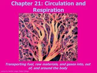
Circulation and Respiration
480 likes | 505 Vues
Circulation and Respiration. Gastrovascular Systems. Body plan is only two or a few cell layers thick. Food enters the same opening as waste exits.

Circulation and Respiration
E N D
Presentation Transcript
Gastrovascular Systems • Body plan is only two or a few cell layers thick. • Food enters the same opening as waste exits. • The fluid in the cavity of cnidarians is continuous with that around them and Planarians have a branching cavity that is adequate for their small thin (flat) bodies.
Larger animals • Need to have a Circulatory system because diffusion takes too long. • A system has blood, a heart/pump that generates blood pressure. • Circulatory systems are powered by cellular energy • Open systems – blood is not separated from body fluid, it bathes the organs, this “blood” is called hemolymph. Blood is pumped by the heart in to spaces called sinuses. It flows back to the heart via a pressure gradient
Closed systems • Blood is confined to vessels and maintained separate from the body fluid. • Cardiovascular systems consist of closed system with a heart the has one or two ventricle and one or two atria
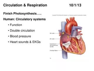
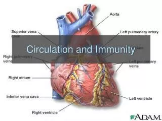
Cardiovascular Systems • Atria – heart chambers that receive blood • Ventricles – heart chambers that pump blood • Artery – vessel that carries blood away from the heart. • Arterioles – small vessels that carry blood to the capillaries • Capillaries(bed) thin walled vessel that infiltrate the body, material exchange occurs here. • Venules – small vessels that carry blood to veins • Veins – carry blood to the heart
Pulmocutaneous circulation • Amphibians have vessels leading from the pulmonary arteries to the skin that receive deoxygenated blood and allow the capillaries in the skin to remove carbon dioxide and even absorb some oxygen.
Systemic and Pulmonary Circulation • Systemic - Supplies nourishment and removes wastes from the tissue located throughout the body • Pulmonary – blood vessels in the lungs flow to and from capillaries where oxygen is absorbed and carbon dioxide is removed.
Double Circulation (not found in fish) • Blood flowing through the lungs is pumped separately from the blood flowing to the body. • Much stronger flow to the brain, muscles, ect.
ECG or EKG • Electrocardiogram measures the electrical currents that are conducted in the body as the heart goes through the cardiac cycle
Movement of blood • Adam Movement of blood overview • Movement of oxygen and Carbon Dioxide
capillaries • Capillary function • Pressure – fluid loss and regain
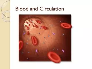
V. On outline - Blood • Blood cells and cell fragments occupy about 45% of the blood volume. • 55% is plasma. • Plasma is 90% water, it contains electrolytes. • Plasma proteins help to maintain pH, osmotic balance, and blood viscosity. • Some of these proteins are immunoglobulins that function in defense.
Cellular Elements • Blood plasma suspends 3 elements: • 1. RBC’s--oxygen transport, most numerous.(eurythrocytes) • 2. WBC’s--defense of body.(leukocytes) • 3. Platelets--fragments of cells which help in the clotting process.
Eurythrocytes • Shape is related to its function. • Biconcave increases its surface area. • Small size and number increases surface area--related to function. • Mammalian lack nuclei--allows for more hemoglobin.
Leukocytes • These are white blood cells and there are 5 types: • 1. Monocytes, Neutrophils, Basophils, Eosinophils, Lymphocytes • Collectively, these fight infection. • These spend most of their time in the interstitial fluid where they fight invaders.
Platelets • These plug wounds and prevent blood loss. • Wounds release factors that make platelets sticky and enable them to adhere to collagen fibers in connective tissue slowing blood loss.
Stem cells • In the marrow of some bones, particularly the ribs, vertebrae, breastbone and pelvis the blood cells are created from pluripotent cells that can produce.
Blood clotting • Platelets plug wounds and prevent blood loss. • Wounds release factors that make platelets sticky and enable them to adhere to collagen fibers in connective tissue slowing blood loss.
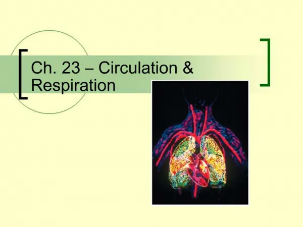
Respiratory Systems • Respiratory surfaces allow for the exchange of gases. • They are always thin and bathed in water. • In most animals, the respiratory medium is a thin, moist epithelium. • This separates the respiratory medium from the blood.
Gills • Are out-foldings of the body surface suspended in water. • They are loaded with capillaries. • Animals with gills ventilate them which moves water with a high concentration of O2 over them.
Tracheal System • Found in insects. • It is made up of tubes that branch through the body which is a variation on a folded, internal respiratory surface. • The trachea branches smaller and smaller and contacts nearly every cell.
Lungs • These are respiratory organs found in one spot of the body. • They have a dense net of capillaries immediately below the epithelium on the respiratory surface. • They are connected to a closed system that transports gases to and from other regions of the body.
Ventilating the lungs • Positive pressure breathing-amphibians • Negative pressure breathing-humans • Diaphragm – muscle below rib cage • Tidal volume is the volume of air inhaled with each breath. • Max. during forced breathing is 3-4.8L • Residual volume is the amount remaining in the lungs after a forced exhale.
Vital capacity • maximum volume of air that a person can exhale after maximum inhalation. It can also be the maximum volume of air that a person can inhale after maximum exhalation.
Control centers in the brain • Human breathing is mostly under autonomic control. • 2 regions of the brain control this: • The pons and the medulla. • The pons controls the medulla which sets a basic breathing rhythm.
Sensors in the aorta and carotid arteries exert secondary control over breathing. • These sensors monitor O2, CO2 and blood pH. • The pH is largely controlled by CO2 levels.
Gases • When CO2 levels increase, carbonic acid levels increase lowering the blood pH. • When pH drops, the depth and rate of breathing increases helping to remove excess CO2. • O2 levels only have an effect on breathing rate at high altitudes.
More Carbon Dioxide • In addition to transporting O2, hemoglobin helps transport CO2 and assists in buffering. • Respiring cells produce CO2. Carbonic anhydrase catalyzes the reaction of CO2 with H2O to form H2CO3. • H2CO3 dissociates into H+ + HCO3- • Most of the H+ attaches to hemoglobin and other proteins minimizing the change in blood pH.
Diffusion in the Alveoli Deoxygenated Blood Air In & Out CO2 O2 Alveolus Blood Capillary Oxygenated Blood
Gas Exchange at the Cells Body Cells CO2 O2 Blood Capillary Tissue Fluid