Download
1 / 27
370 likes | 1.3k Vues
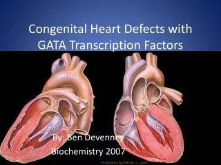
OntoDiagram Ontology Based Diagram Generation Congenital Heart Defects in Pediatric Cardiology. Kartik Vishwanath, Venkatesh Viswanath, Yugyung Lee, Ph.D School of Computing and Engineering University of Missouri—Kansas City William Drake, MD Children's’ Mercy Hospital—Kansas City.

E N D
OntoDiagramOntology Based Diagram GenerationCongenital Heart Defects in Pediatric Cardiology Kartik Vishwanath, Venkatesh Viswanath, Yugyung Lee, Ph.D School of Computing and Engineering University of Missouri—Kansas City William Drake, MD Children's’ Mercy Hospital—Kansas City
OntoDiagram Application for pediatric cardiology that generates Mullin’s like diagram based on domain descriptions, clinical observations and measurements • Mullin’s Atlas—A collection of widely used diagrammatic representation of anatomy of heart structure (Mullins Diagram) • Mullin’s Diagrams are useful in hospital reporting tools as they efficiently represent the defects • Given a description of heart by domain experts, the system should automatically generate the Mullin’s diagram representing the conditions as described
Sample Mullin’s Diagrams Pulmonary Atresia Tricuspid Atresia Complex TOF TAPVR LSVC
Comprehending textual descriptions is time consuming! Situs and Relations: There is levocardia with visceral and atrial situs solitus, atrioventricular concordance (D-looped ventricles) and normally related great arteries {S,D,S} noteVenous Connections: There are normal systemic and pulmonary venous connections, with the superior and inferior vana cavae returning normally to the right atrium, and all four pulmonary identified returning normally to the left atrium. noteAtria: The left and right atria have normal chamber size, structure, and relations. noteAtrioventricular Valves: The mitral and tricuspid valves have normal structure, size, placement and function. There is no mitral insufficiency and only physiologic tricuspid insufficiency. noteVentricles: There is normal left ventricular chamber size and normal subjective left ventricular funtion with flattened interventricular septal motion. note There is normal right ventricular chamber size. note The right ventricle wall thickness is moderately increased. note There is normal global left ventricular function. note There is flattened interventricular septal motion. noteOutflow Tracts: The left ventricular outflow tract has normal size and geometry, without stenosis or narrowing. note The right ventricular outflow tract has moderate hypoplasia. note There is no subaortic conus. note There is marked right ventricular outflow tract turbulence (stenosis). note The peak right ventricular outflow tract velocity is 4.1 meters/second. note The peak right ventricular outflow tract gradient is 67 mmHg. noteSemilunar Valves: The aortic valve is normal, with three normal leaflets, normal mobility and no prolapse. note There is non-turbulent flow and a normal flow velocity across the aortic valve with no evidence of stenosis. note There is marked hypoplasia of the pulmonary valve annulus. note The pulmonary valve annulus measures 2.1 mm in the long axis outflow view. note The peak velocity across the aortic valve is 1.3 meters/second. note The pulmonary valve leaflets are moderately thickened. noteAortic Root Ascending Aorta: The aortic root, including the sinuses of valsalva, sinotubular ridge, and proximal ascending aorta are normal without stenosis, narrowing, or dilation. notePulmonary and Thoracic Arteries: There was supravalvular pulmonary stenosis present. note There was hypoplasia of the main pulmonary artery. note The left pulmonary artery appeared hypoplastic. note The right pulmonary artery appeared hypoplastic. note There was a right-sided aortic arch present. note There was a patent ductus arteriosus visualized. note There was a right-sided patent ductus arteriosus present. note There was left-to-right shunting demonstrated across the patent ductus arteriosus. note There was a right aortic arch with a retroesophageal segment and left descending aorta forming a vascular ring. noteSeptal Defects: There is a malalignment type ventricular septal defect present. note The ventricular septal defect measures 12 mm from the apical four-chamber view. note There is a moderate degree of aortic override. Fibro-annular continuity of the aortic and mitral valves is present. note The aorta has about 50 per cent override. note
Diagram is easier to understand! Complex TOF
Motivation • Visual representation is intuitively easier to understand than text information. • Pediatric Cardiology faces new or variations of heart defects • Mullin’s atlas are comprehensive but not complete • Difficult to search and retrieve similar diagrams • Domain experts manually draw diagrams by hand or modify existing ones in Mullin's atlas
Domain World Patent Foramen Ovale Pulmonary Artery Right-left shunt Coarctation anterior Bifurcation of PA True PA Aortic stenosis Normal Bicuspid valve Parachute Mitral Valve PDA Tricuspid insufficiency Domain Experts’ Perspective Overriding Aorta hypoplasia Ontological Framework Mitral valve cleft Heterogeneity of Perspectives above Hidden behind translation translation below transposition Diagram Perspective Component sheared Scaled along long axis connections Diagram World missing
Challenges • Heart Structure—”simple & complex” • Mapping domain knowledge onto diagram specifications • Perspective and representational gap between medical domain and diagram domain • Domain Modeling • Modeling congenital defect information • Modeling anatomical structure • Implicit domain knowledge • Anatomical consequence of defects • Diagram Modeling • Individual component modeling • Spatial orientation between components • Transformations on components to reflect changes
Heart Structure“Simple &Complex” Simple • An anatomically simple structure • Intuitively hierarchical “part-of” relationships (left heart, right heart, etc.) • Heart componentsare less in count • Anatomically new additions are very less and predictable Complex • Variations of each component are large • Variations of heart as a whole are diverse • Complex relationship between various anomalous conditions
OntoDiagram: Focus • Translation from domain description to diagram • Key issues • Domain modeling (Ontology approach) • Diagram modeling (Component based approach) • Mapping domainonto diagram (Domain rules) OntoDiagram Physician’s description Diagram
OntoDiagram Architecture Congenital Heart Defect Ontology Heart Anatomy Ontology Measurement Ontology Diagram Ontology DomainOntologies Diagram Ontology Mapping System OntoDiagram Query Interface Domain Rules Diagram repository Databases Diagram Composer Medical Database Medical Database
Ontologies • An ontology is an explicit formal specification of the terms in the domain and relations among them (Gruber 1993). • To share common understanding of the domain knowledge on congenital heart defects among cardiologists, nurse, database engineer or software agents • To make domain assumptions explicit so that defect associations patterns could be extracted • To separate domain knowledge from operational knowledge • To map different perspectives of domain and diagram
Domain Modeling • Modularizing domain knowledge • Congenital Heart Defect Ontology [UMLS] • Classification of defects, anomalous conditions • Definition for domain terms like stenosis, atresia, etc. • Anatomical consequences of defects • Association between defects • Anatomy ontology [FMA] • Multi-perspective classification of heart anatomy • Possible anatomical changes in each heart component • Orientation between components • Measurement and Diagram Ontology [LOINC]
Diagram Modeling • Diagram Models • Component Association Model (Spatial Orientation between components) • Structure Model (Component hierarchy) • Conversion Model (Transformations) • Annotation Model (Relevant annotations) • Diagram perspective of changes • Abnormal growth • Missing parts • Transposition • Transformation • Combination
right right top top top right top bottom Component Association Model Spatial relations with components and their neighboring components • Six-tuple {Ct, Cn, D, A, O, P} where • Ct and Cn components, D—spatial orientation of Ct with respect to Cn, A—interface presence, O—orientation, P—presence of component e.g. {Descending Aorta, Aortic Arch, bottom, attached, below, present}
Structure Model • The heart is considered to be made of two layers • The first layer consisting of the chambers of the heart (wall of atria and ventricles) • The second layer consists of the other components of heart (Pulmonary artery and Pulmonary valves, Aorta and Aorta valves, etc.) • The relationships between the first and the second layer components
Interface Points & Gate Points Layer—Layer Component—Component
Conversion Model • Transformation of components • Geometric transformation (e.g., Scale, Rotate, Translate) • Polynomial transformation (e.g., Shear) • Domain specific transformation (e.g, dilation, coarctation) • Transformation model defined as {CT,T, P} • CT—component being transformed • T—name of the transformation • P—transformation parameters
Hierarchical Composition • Hierarchical model—closer to domain perspective • Faster diagram generation—few components change • Optimal selection of components
Hierarchical Composition Steps Step 1: Configuration • Identify an appropriate set of components and their abstraction level to be composed [Congenital Defects and Association models] • Determine the components to be transformed [Conversion Model] • Determine the relationships between components (below, above) [Component Structure Model] Step 2: Composition Starting from the most specific level to the root • Transform component images using the transformation operations if necessary (scale, rotation, etc) • Rank the components [Image Model - Color Schema] • Compose the components using interface points [Structure Model] • Forward the interface points to the upper level Step 3: Annotation
OntoDiagram System Ontological Framework Congenital Heart Defect Ontology Anatomy Ontology Domain ontologies Measurement Ontology Diagram Instruction Files MappingSystem Domain Description Diagram Ontology Diagram Composer
OntoDiagram Prototype • Constructed domain ontologies using the protégé in OWL. • Implemented the query interface in Java using the Jena. • Implemented the image composition module using the Java Advanced Imaging package.
Sample Output (Patent Ductus Arteriosus)
Conclusion • The domain descriptions of the congenital heart defects are mapped to generate a diagrammatic representation of defects. • Ontology based domain modeling • Component based diagram modeling • Rule based mapping between domain and diagram • Hierarchical composition of components • A prototype system is currently under testing at the Children’s Mercy Hospitals and Clinics • facilitate existing treatments for managing patients with severe heart disorders.