Hepatitis B Virus
Hepatitis B Virus. Christian A. García Sepúlveda MD PhD. Laboratorio de Biología Molecular Facultad de Medicina Universidad Autónoma de San Luis Potosí. Hepatitis B Virus Introduction. 250 million people infected worldwide.


Hepatitis B Virus
E N D
Presentation Transcript
Hepatitis B Virus Christian A. García Sepúlveda MD PhD Laboratorio de Biología Molecular Facultad de Medicina Universidad Autónoma de San Luis Potosí
Hepatitis B VirusIntroduction • 250 million people infected worldwide. • In areas of Africa and East Asia, 50% of the population may be seropositive, 5-15% may be chronically infected (carriers). • Carriers are 200x more likely than non-carriers to develop primary hepatocellular carcinoma. • 300,000 cases per year in the US; 4,000 fatalities. • 70-90% of maternal-neonatal infections result in chronic infection. ³8% - High: Early childhood infection, lifetime risk of infection 60% 2-7% - Intermediate: Infection at all ages, lifetime risk of infection 20%-60% <2% - Low: Infection as adult, lifetime risk of infection <20%
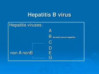
Hepatitis B VirusTaxonomy Hepadnavirus family has primate (HBV) as well as Rodent (WHV) and avian (DHBV) representatives. HBV and WHV have 80% homology (nt) HBV and DHBV have 40% homology (nt) Hepadnavirus must have existed before the speciation of birds and mammals. DHBV possesses the smallest genome of known animal viruses (3021 bp).
Hepatitis B VirusTaxonomy Baltimore Classification: Group VII for dsDNA-RT integrating viruses. Enveloped virion containing partial double-stranded circular DNA genome Replication occurs through an RNA intermediate Virus encodes and carries a reverse transcriptase Virus encodes several antigenically and clinically predictive important proteins
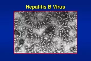
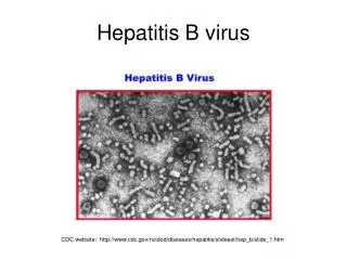
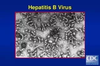
Hepatitis B VirusMorphology Virion also referred to as Dane particle. 42 nm enveloped virus. Expresses surface antigens (HBsAg). Core antigen (HbcAg) located in the center (nucleocapsid). Minor core component e antigen (HBeAg) antigenically distinct from HBcAg. 22 nm spheres and filaments are not infectious, outnumber the actual virions (subviral). 25 nm 22 nm 100 – 700 nm HBsAg = 4 phenotypes : adw, adr, ayw and ayr HBcAg = inner core protein (a single serotype) HBeAg = secreted protein; function unknown
Hepatitis B VirusMorphology Outer plasma membrane derived from host cell (envelope) Surface antigens embedded in envelope are glycoproteins. Three sizes (L, M & S) glycoproteins. Icosahedral core assures stability (HBcAg)
Hepatitis B VirusMorphology Three Surface Glycoproteins: The small protein (SHBs) is encoded by the S region. - 80-90% of Surface GP. Large protein (LHBs) encoded by pre-S1, pre-S2, and S region. - 1-2% of Surface GP. Middle protein (MHBs) encoded by the pre-S2 and S regions. - 5-15% of Surface GP. The SHBs is the most common form of these proteins.
Hepatitis B VirusGenome “Dane particles” enclose a single circular, incomplete double stranded DNA genome. Regulatory and structural sequences for viral transcription included. Extensive overlap of genes. HBV genome adopts two forms: rc-DNA and cccDNA In the virion, the genome exists as rcDNA (transcriptionally useless but great at saving information). In the hepatocyte, the genome exists as cccDNA which is transcriptionally active and serves as a reference to replicate more rcDNA. rcDNA cccDNA
Hepatitis B VirusGenome rcDNA Relaxed circular DNA Partially double-stranded L strand or (-)DNA strand is complete and has a short 5’ terminal redundancy covalently coupled to a protein (polymerase). S strand or (+)DNA strand is incomplete, only 2/3 of the L strand, 5’ end has RNA primer and the length of the 3’ end is variable. 3’ end of L strand overlaps 5’ end of S strand. rcDNA cccDNA
Hepatitis B VirusGenome cccDNA Covalently Closed Circular DNA After cell entry (+)DNA strand is completed. 5’ RNA oligo is removed from the (+)DNA. Pol and 3’ short terminal redundant sequences are removed from (-)DNA. Two strands are covalently ligated by host repair enzymes. Strands are stabilized with host cell nucleosomes and might be supercoiled. Remains as a plasmid within hepatocytes. Used as a replication/transcription template. rcDNA cccDNA
Hepatitis B VirusGenome The partially double-stranded relaxed circular DNA (rcDNA) indicated with thick black line. P covalently linked to the 5´ end of the (-)DNA. RNA primer (zigzag line) at the 5´ end of (+)DNA. The dashed line symbolizes the heterogeneous lengths of the (+)-strands. DR1 and DR2 are the direct repeats. The outer circle symbolizes the terminally redundant pgRNA with ε close to the 5´ end, and the poly-A tail at the 3´ end. The precore mRNAis nearly identical, except it starts slightly upstream. The relative positions of the open reading frames for core (C), P, preS/S, and X are shown inside.
Hepatitis B VirusGenome The partially double-stranded relaxed circular DNA (rcDNA) indicated with thick black line. P covalently linked to the 5´ end of the (-)DNA. RNA primer (zigzag line) at the 5´ end of (+)DNA. The dashed line symbolizes the heterogeneous lengths of the (+)-strands. DR1 and DR2 are the direct repeats. The outer circle symbolizes the terminally redundant pgRNA with ε close to the 5´ end, and the poly-A tail at the 3´ end. The precore mRNAis nearly identical, except it starts slightly upstream. The relative positions of the open reading frames for core (C), P, preS/S, and X are shown inside.
Hepatitis B VirusGenome The partially double-stranded relaxed circular DNA (rcDNA) indicated with thick black line. P covalently linked to the 5´ end of the (-)DNA. RNA primer (zigzag line) at the 5´ end of (+)DNA. The dashed line symbolizes the heterogeneous lengths of the (+)-strands. DR1 and DR2 are the direct repeats. The outer circle symbolizes the terminally redundant pgRNA with ε close to the 5´ end, and the poly-A tail at the 3´ end. The precore mRNAis nearly identical, except it starts slightly upstream. The relative positions of the open reading frames for core (C), P, preS/S, and X are shown inside.
Hepatitis B VirusGenome The partially double-stranded relaxed circular DNA (rcDNA) indicated with thick black line. P covalently linked to the 5´ end of the (-)DNA. RNA primer (zigzag line) at the 5´ end of (+)DNA. The dashed line symbolizes the heterogeneous lengths of the (+)-strands. DR1 and DR2 are the direct repeats. The outer circle symbolizes the terminally redundant pgRNA with ε close to the 5´ end, and the poly-A tail at the 3´ end. The precore mRNAis nearly identical, except it starts slightly upstream. The relative positions of the open reading frames for core (C), P, preS/S, and X are shown inside.
Hepatitis B VirusGenome The partially double-stranded relaxed circular DNA (rcDNA) indicated with thick black line. P covalently linked to the 5´ end of the (-)DNA. RNA primer (zigzag line) at the 5´ end of (+)DNA. The dashed line symbolizes the heterogeneous lengths of the (+)-strands. DR1 and DR2 are the direct repeats. The outer circle symbolizes the terminally redundant pgRNA with ε close to the 5´ end, and the poly-A tail at the 3´ end. The precore mRNAis nearly identical, except it starts slightly upstream. The relative positions of the open reading frames for core (C), P, preS/S, and X are shown inside.
Hepatitis B VirusGenome The partially double-stranded relaxed circular DNA (rcDNA) indicated with thick black line. P covalently linked to the 5´ end of the (-)DNA. RNA primer (zigzag line) at the 5´ end of (+)DNA. The dashed line symbolizes the heterogeneous lengths of the (+)-strands. DR1 and DR2 are the direct repeats. The outer circle symbolizes the terminally redundant pgRNA with ε close to the 5´ end, and the poly-A tail at the 3´ end. The precore mRNAis nearly identical, except it starts slightly upstream. The relative positions of the open reading frames for core (C), P, preS/S, and X are shown inside.
Hepatitis B VirusGenome The partially double-stranded relaxed circular DNA (rcDNA) indicated with thick black line. P covalently linked to the 5´ end of the (-)DNA. RNA primer (zigzag line) at the 5´ end of (+)DNA. The dashed line symbolizes the heterogeneous lengths of the (+)-strands. DR1 and DR2 are the direct repeats. The outer circle symbolizes the terminally redundant pgRNA with ε close to the 5´ end, and the poly-A tail at the 3´ end. The precore mRNAis nearly identical, except it starts slightly upstream. The relative positions of the open reading frames for core (C), P, preS/S, and X are shown inside.
Hepatitis B VirusGenome The partially double-stranded relaxed circular DNA (rcDNA) indicated with thick black line. P covalently linked to the 5´ end of the (-)DNA. RNA primer (zigzag line) at the 5´ end of (+)DNA. The dashed line symbolizes the heterogeneous lengths of the (+)-strands. DR1 and DR2 are the direct repeats. The outer circle symbolizes the terminally redundant pgRNA with ε close to the 5´ end, and the poly-A tail at the 3´ end. The precore mRNAis nearly identical, except it starts slightly upstream. The relative positions of the open reading frames for core (C), P, preS/S, and X are shown inside.
Hepatitis B VirusOpen Reading Frames (ORFs) • There are 4 open reading frames in the same strand (+)DNA • S– the surface antigen • Three different polypeptides producedfrom three different alternative translation ATGs. • C - the core protein • And its minor (preC) component • P - the polymerase • Has a Terminal Protein (TP) domain. • X– the viral transcription transactivator. • Conserved in all mammals (but not avian) hepadnaviruses. • Though not essential in transfected cells, it is required for infection in vivo.
Hepatitis B VirusOpen Reading Frames (ORFs) HBV Genome has an inner (+)DNA strand that is almost circular and has varying lengths. (+)DNA strand has a DR2 and DR1 cassette at the 5’ end.
Hepatitis B VirusOpen Reading Frames (ORFs) An outer (-)DNA strand is full length and close to 3.5 kb long. It also has a DR1 at the 5’ end and a DR2 at the 3’ end. (-)DNA DR1 and DR2 complement (+)DNA DR1 and DR2. It is covalently associated to a Terminal Protein Domain of the Polymerase.
Hepatitis B VirusOpen Reading Frames (ORFs) Most of the HBV genome is dedicated to encoding the Polymerase (ORF P). Surface antigen (S) is encoded as a single mRNA transcript which includes: - pre-S1 - pre-S2 and - ORF S Alternate start codons lead to their translation. Core protein and minor component ara encoded by first ORF (ORF C). Last ORF codes for X protein (ORF X).
Hepatitis B VirusOpen Reading Frames (ORFs) Each ORF is transcribed into seperate mRNAs. Each mRNA is translated to produce the different proteins.
Hepatitis B VirusReplication • Replication of the hepadnaviral genome can broadly be divided into three phases: • (1) Infectious virions contain a partially double-stranded circular but not covalently closed DNA genome of about 3.2 kb in length (relaxed circular, or rcDNA) inside their inner icosahedric core.
Hepatitis B VirusReplication • (2) Upon infection, the RC-DNA is converted, inside the host cell nucleus, into a plasmid-like covalently closed circular DNA (cccDNA).
Hepatitis B VirusReplication • (3) Genomic and subgenomic RNAs are transcribed by cellular RNA polymerase II from cccDNA.
Hepatitis B VirusReplication • - Pregenomic RNA (pgRNA) selectively packaged into progeny capsids and is reverse transcribed by the P protein into new rcDNA genomes.
Hepatitis B VirusReplication • - Matured rcDNA containing-but not immature RNA containing nucleocapsids can be used for intracellular cccDNA amplification, or be enveloped and released from the cell as progeny virions.
Hepatitis B VirusGenotypes First DNA sequence for HBV publshed in 1979 by Galibert et al. Genotypes are defined as a group of nt sequences that exhibit ≥ 92% homology and diverge from other sequences by ≥ 8%. Genotypes geographical distribution A & D relatively ubiquitous. D rare in N Europe & Americas. G is least known, posibly ubiquitous. B & C are predominanty Asian E Subsaharan Africa F South/Central America H Central America and Southern US Phylogenetic tree of HBV genotypes and subtypes.
Hepatitis B VirusGenotypes First DNA sequence for HBV publshed in 1979 by Galibert et al. Genotypes are defined as a group of nt sequences that exhibit ≥ 92% homology and diverge from other sequences by ≥ 8%. New World Genotypes F & H are on same phylogenetic branch as the WM Non-human Primate WM sequences… Indictaes a zoonotic transmission of these genotypes to humans of Central and South America. Phylogenetic tree of HBV genotypes and subtypes.
Hepatitis B VirusGenotypes Characteristics of HBV genotypes and subtypes Main serotypes in bold bp aminoacids
Hepatitis B VirusGenotypes and Clinical Outcome A genotypes may lead to more Chronic Hepatitis than D genotypes in some countries. A genotypes also easier to clear. C genotypes progress more rapidly to cirrhosis and hepatocelular carcinoma. F genotypes (Amazonian basin) linked to fulminant hepatitis in HDV coinfected Hosa. G genotypes associated with increased liver fibrosis in HIV patients. Genotypes A & B have higher chance of becoming HBeAg negative than Genotypes C & D. In the Japanese population, Genotype B and C respond better to interferon treatment in contrast to Genotype A patients.
Hepatitis B VirusEmergence of Viral Mutants Mutations can arise in any part of the
Hepatitis B VirusSerologic Course (Acute Infection w/recovery) HBsAg Can be detected in high levels in serum during acute or chronic HBV infection. The presence of HBsAg indicates that the person is infectious. The body normally produces antibodies to HBsAg as part of the normal immune response to infection. HBsAg is the antigen used to make hepatitis B vaccine.
Hepatitis B VirusSerologic Course (Acute Infection w/recovery) Anti-HBs The presence of anti-HBs is generally interpreted as indicating recovery and immunity from HBV infection. Anti-HBs also develops in a person who has been successfully vaccinated against hepatitis B. HBsAg is the antigen used to make hepatitis B vaccine.
Hepatitis B VirusSerologic Course (Acute Infection w/recovery) Total anti-HBc Total hepatitis B core antibody. Appears at the onset of symptoms in acute hepatitis B and persists for life. The presence of anti-HBc indicates previous or ongoing infection with HBV in an undefined time frame.
Hepatitis B VirusSerologic Course (Acute Infection w/recovery) IgM anti-HBc IgM antibody to hepatitis B core antigen. Positivity indicates recent infection with HBV (≤6 months). Its presence indicates acute infection.
Hepatitis B VirusSerologic Course (Acute Infection w/recovery)
Hepatitis B VirusSerologic Course (Acute Infection w/recovery)
Hepatitis B VirusSerologic Course (Acute Infection w/recovery)
Hepatitis B VirusSerologic Course (Acute Infection w/recovery)
Hepatitis B VirusSerologic Course (Acute Infection w/recovery)
Hepatitis B VirusSerologic Course (Acute Infection w/recovery)
Hepatitis B VirusHBV Testing Testing recommended for: - pregnant women - infants born to HBsAg-positive mothers - household contacts and sex partners of HBV-infected persons - healthcare workers, laboratory personel - persons infected with HIV Serologic testing for hepatitis B surface antigen (HBsAg) is the primary way to identify persons with chronic infection with HBV infection (CDC, Sep 2008).
Hepatitis B VirusHBV Testing (ELISA) • Advantages • 96 tests format • Objective results • Automatable • Appreciable sensitivity & Specificity • Narrower detection window • Disadvantages • Demands skill sets • Decade old method • Detection capability surpassed by newer methods • Sero-conversion detection panels