Download
1 / 84
910 likes | 1.12k Vues
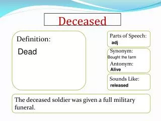
Hemodynamics of the Neurologically Deceased Donor. For the Donation and Transplantation Community of Practice August 4, 2014 Webcast. Planning Committee Members. Daniel J. Lebovitz, MD

E N D
Hemodynamics of the Neurologically Deceased Donor For the Donation and Transplantation Community of Practice August 4, 2014 Webcast
Planning Committee Members • Daniel J. Lebovitz, MD Associate Medical Director, Division of Critical Care, Akron Children’s Hospital, Director PICU, MetroHealth Medical Center, Medical Director, Lifebanc • LeAnn Swanson, MPH Executive Director, Organ Donation and Transplantation Alliance • Teresa Beigay, DrPH Director of Special Initiatives, HHS/HRSA/HSB/Division of Transplantation • Roxane Cauwels, BSN, MBA DTCP Consultant, Organ Donation and Transplantation Alliance
Presenters • Daniel J. Lebovitz, MD Associate Medical Director, Division of Critical Care, Akron Children’s Hospital, Director PICU, MetroHealth Medical Center, Medical Director, Lifebanc • Anil S. Paramesh, MD, FACS Associate Professor of Surgery, Urology and Pediatrics, Director, Living Donor Transplant Program, Tulane Abdominal Transplant Institute, Tulane University School of Medicine, Clinical Associate Professor of Surgery, Louisiana State University Health Sciences Center, AOPO Medical Director • Harry E. Wilkins, III, MD, MHCM, FACS Trauma Surgery & Surgical Critical Care, Quincy Medical Group • Raghavan Murugan, MD, MS, FRCP, FCCP Associate Professor of Critical Care Medicine and Clinical and Translational Science, Core Faculty, Center for Critical Care Nephrology, CRIMSA Center, Department of Critical Care Medicine, University of Pittsburgh
Objectives • To provide information on the hemodynamic derangements of brain death • To improve understanding of minimally invasive and non-invasive hemodynamic monitors • To share the results of the MOnIToR study • To integrate effective organ donor management principles and practices into the continuum of end-of-life hospital care • To encourage critical and palliative care physicians and staff to be actively engaged in the organ donation process for preserving the option of donation for all families
US Cadaveric Donors, Cadaveric Transplants,and Number on Waiting List (1990-present) www.optn.transplant.hrsa.gov Waiting List July 30, 2014: 123,115 Transplants Donors Deaths on the list
Goals in the Management of an Organ Donor • Maximize the number of organs transplanted per donor • Maximize the function of the transplanted organ in the recipient
What gets in our way? • The pathophysiology of Brain Death • Hemodynamic instability • Pulmonary compromise • Oxygen delivery to hypoxic/ ischemic organs
Key Goal in the Management of the Organ Donor • Optimize cardio-respiratory function to achieve and maintain adequate organ perfusion without volume overloading • Re-establish and/or maintain adequate Oxygen Deliver (DO2) to organs that may be transplanted
Outline of Today’s Webcast • Discussion of hemodynamic physiologic derangements of the neurologically deceased donor • Tools of the Trade – Hemodynamic Monitoring • Any data on using these tools in Organ Donor Management? • Wrap Up and Question Session
Cardiovascular Physiology of the Brain Dead Donor Anil S. Paramesh, MD, FACS Associate Professor of Surgery, Urology & Pediatrics Tulane University School of Medicine Medical Advisor, AOPO
Brain Death • Brain Death is associated with widespread physiologic changes that cross multiple organ systems and can lead to hemodynamic compromise
Severe Brain Injury Neurologic Manifestations Electrolyte Disorders Endocrine Abnormalities Pulmonary Injury Hypothermia Coagulopathy
The Progression of Brain Death Characterized by progression of ischemia from head downwards Cerebrum Brainstem Spinal Cord
Cerebral Ischemia • During the process of herniation, body tries to preserve cerebral blood flow • Stimulation of vagal and sympathetic systems • Cushing’s triad – hypertension, bradycardia and abnormal breathing
Brain Stem Ischemia • Necrosis of nerve cells in the medulla causes unchallenged sympathetic activation • “Autonomic storm” - 800 to 1200 times increase in catecholamine levels in blood • Increased SVR, CO, heart rate, MAP, cardiac O2 demand • Can cause myocardial ischemia and/or arrhythmias
Spinal Ischemia • May occur minutes to hours after Storm • Loss of sympathetic overdrive • Vasodilation, decreased BP, HR, contractility • This stage represents greatest risk to donor organs
Volume Capacities of Brain Death Catecholamine squeeze Mannitol Blood loss DI Brain death 10 liters 10 liters 5 liters 7 liters
Hormonal effects of Brain Death • Decrease in ADH, T3, cortisol and insulin • Electrolyte disorders • ADH leads to DI – massive fluid losses • T3 • Switch in cardiac cells to anaerobic metabolism • Increased lactate and acidosis • Myocardial dysfunction
Hypothermia • Loss of function of hypothalamus • Contributes to hemodynamic instability → Myocardial depression/arrythmias → Reduced tissue oxygenation → Impaired kidney function
Coagulopathy • Release of tissue thromboplastin and plasminogen from necrotic brain tissue • Hypothermia and dilution also contribute • May contribute to worsening bleeding
Pulmonary Flow • During the Storm phase • Increase in LAP - decreased LV output due to severe SVR • Temporary cessation of pulmonary blood flow with pressures exceeding hydrostatic pressures • Neurogenic pulmonary edema and hemorrhage • Impairment LV > RV - CVP may not be accurate measurement of LV function
Electrolyte Imbalances • Hypernatremia • Hyper/Hypokalemia • Hypocalcemia • Hypo/Hyperglycemia
Brain Death Diabetes insipidus Dehydration Hormone depletion Loss of vasomotor tone Anaerobic metabolism Less Preload Less afterload Less coronary perf Less Ca+ channel stimulation Electrolyte disorders CV dysfunction
Minimally Invasive and Non-Invasive Hemodynamic Monitors Harry E. Wilkins, III, MD, MHCM, FACS and Daniel J. Lebovitz, MD
What do we need to know? • Intravascular fluid status of the donor • Reliable metrics / goals to guide resuscitation • How to initiate the monitoring
First, a little background… • Early in the Collaborative, MTN striving to meet 3.75 OTPD • Critical Care Task Force convened – 2005 • Revisited and revised Donor Management Goals with aggressive management of Donors in mind • No local lung program and poor thoracic organ yield • Suspected we were losing donors to less than optimal management
Case Illustration 59FC Involved in MVC 2 weeks prior Brief LOC at the scene No acute head injury on CT but found two unruptured aneurysm Referred for NS consult Considered stable and sent home to await NS appointment
Case Illustration, cont. Found the next week unresponsive CT revealed large ICH Not a candidate for surgical intervention Admitted to ICU for supportive care 18 hours later declared Brain Death PMH: HTN on meds for one year ½-1PPD smoker for 39 years active horse rider/employed on ranch
What we ‘need’ Maximum data regarding hemodynamic status of our patient population Cardiac performance (C.O./ C.I.; SV) Vascular tone (Systemic Vascular Resistance) Intravascular volume Portability Ease of application / use Accuracy, reliability, reproducibility
Clinical Exam Blood pressure cuff Central Venous Pressure readings Pulmonary Artery (Swan Ganz) Catheter Serial Lactate Measurements Flotrac LiDCO PiCCO Inadequate Very basic and limited Only right-sided pressures and shown to be inaccurate Lots of good data, yet falling out of favor / use ‘Lagging’ data Art. Waveform analysis Indicator dilution Pulse contour analysis Our Options
Clinical Exam • Inspection • Skin turgor • Urine output • Cardiac auscultation • Peripheral pulse characteristics • Straight leg raising test • Orthostatics
Central Venous Pressure • Right sided pressures, only • Requires interpretation of waveform with respect to respiratory cycles and can be effected by interpretation • Readings may be affected by other conditions that affect thoracic pressures (PEEP, pneumothorax, pulmonary HTN, pericardial effusion, etc.) • Gives no information regarding cardiac performance • Recent studies suggest CVP is not reliable in determining fluid responsiveness* *Does central venous pressure predict fluid responsiveness? A systematic review of the literature and the tale of seven mares. Chest. 2008 Jul;134(1):172-8. doi: 10.1378/chest.07-2331
Indicator Dilution Techniques Pulmonary Artery (Swan-Ganz) Catheter LiDCO Catheter
PA (Swan Ganz) Catheter • Stewart-Hamilton Formula (simplified): • CO = amount of injected indicator area of dilution curve (AUC) • Observed in 1897 and used indocyanine green as the indicator dilution method used to measure CO in critically-ill patients until the 1970’s • Thermodilution method adapted the indicator-dilution principle to injectates that cause changes in blood temperature detected downstream • CO is inversely proportional to the mean blood-temperature depression and the duration of transit of cooled blood (i.e. area under the curve - AUC)
PA (Swan Ganz) Catheter • Catheterization of the heart in man with use of a flow-directed balloon-tipped catheter. SwanHJ, GanzW, ForresterJ, MarcusH, DiamondG, ChonetteD. N Engl J Med 1970; 283: 447-51 • A new technique for measurement of cardiac output by thermodilution in man. GanzW, DonosoR, MarcusHS, ForresterJS, SwanHJ. Am J Cardiol 1971; 27:392-6 • Following the introduction of the pulmonary artery (Swan-Ganz) catheter into clinical practice, this ‘single bolus’, thermodilution measurement of CO has been widely accepted as the ‘clinical standard’ for advanced HD monitoring and still considered the ‘gold standard’ against which new technologies are validated and compared
LiDCO Catheter • Utilizes a combination of indicator dilution method like the PA catheter with Lithium as the indicator • The Lithium ‘wash out’ curve provides accurate absolute CO value and is use to then calibrate and correlate continuous CO measurements
Waveform Analysis Flotrac (art waveform analysis) PiCCO (Pulse Contour analysis)
Flotrac • Utilizes Arterial wave form analysis and the phenomenon of pulsus paradoxus • Cardiac Output (CO) • Stroke Volume Variation (SVV) • (If using the Presep catheter, can obtain oxygen delivery and consumption data, as well…)
Systolic press. PP µ SV Diastolic press. Waveform Analysis • Arterial pressure is sampled at 100 Hz • Changes in stroke volume will result in corresponding changes in the pulse pressure • A robust “whole waveform” measure of the pulse pressure is achieved by taking the standard deviation of the sampled points of each beat • sd(AP) µ Pulse Pressure µ Stroke Volume • SV estimates are calculated every 20 sec
Pulse Contour Analysis • Stroke Volume (Pulse pressure proportional to Stroke Volume) • Aortic Compliance (age, sex, morphology, aortic atheromatosis) • Vascular Tone (clinical condition and therapeutic approach)
Transpulmonary thermodilution • Requires central venous access • Requires a femoral arterial line • Injection point: right atrium • Captation point: Femoral artery • Allows calculation of: • Global End diastolic Volume (GEDV) • Intrathoracic Blood Volume (ITBV) • Extravascular Lung Water (EVLW)
Non-Invasive Monitoring Tools in Critical Care • Ultrasound/ Doppler devices • Bioimpedance/ Bioreactance devices
Ultrasound/ Doppler Devices • Uses Ultrasound and Doppler effect to measure cardiac output (Q) 2D echo measures cross sectional area of an annulus x velocity time integral (the Doppler flow profile) to determine SV - flow volume per beat • SV x HR = Q • Single point in time studies – not easily reproducible, very operator dependent
Trans-esophageal Echocardiography • provides information on cardiac contractility, filling, output, valve morphology, and cardiac function • Flow measured CO is determined by pulsed-wave Doppler across cardiac valve or LV outflow tract and assessment of cross sectional area at aortic valve • Requires the Doppler beam be parallel to the blood flow and the cross sectional area measurement be the exact same over time for accuracy • operator dependent skill and knowledge • significant Inter-observer variability
Transesophageal Doppler • Flow measured CO determination in descending aorta • Numerous probes available • ODM II (Abbott), CardioQ/ Medicina TECO (Deltex), HemoSonic 100 (Arrow) • CO calculated from measured aortic blood flow and aortic cross sectional area estimated by nomograms • Likely not valid in BD donors - nomograms assume significant flow to brain • Probes small, position changes easily needs to be continuously adjusted (10-15 % reproducibility)
Transcutaneous Doppler • uses CW (continuous wave) Doppler velocity time integral (VTI) but uses an anthropometric data algorithm for aortic and pulmonary valve diameters to evaluate both R and L sided flow velocities with improving inter-individual reproducibility • USCOM (ultrasonic cardiac output monitor)
Doppler Techniques • Steep learning curves for these modalities • Positioning • Level • Orientation • Operator dependent • Inter-observer variability • Some preliminary experience in OPO world (NEOB) • Preliminary data encouraging