Chapter 1 Basic Anatomy
Chapter 1 Basic Anatomy. Chris Rorden Coordinates Introduction to the nervous system. Multiple choice. What is an example of a common mnemonic? Someone with blue eyes. Someone with odd eyes (one blue, one green). The left hand is controlled by the right side of the brain.

Chapter 1 Basic Anatomy
E N D
Presentation Transcript
Chapter 1 Basic Anatomy • Chris Rorden • Coordinates • Introduction to the nervous system
Multiple choice • What is an example of a common mnemonic? • Someone with blue eyes. • Someone with odd eyes (one blue, one green). • The left hand is controlled by the right side of the brain. • Kings prefer chess over football, generally speaking.
Mnemonics • Mnemonics: tools to aid memory • Kings prefer chess over football generally speaking = Kingdom, phylum, class, order, family, genus, species. • My very easy method, just set up nine planets = Mercury Venus Earth Mars Jupiter Saturn Neptune Pluto • Other methods include loci (imagine a walk with different objects in different locations) or rhymes (one is a bun, two is a shoe…)
Multiple choice • What is the symbol for Sagittarius? • The Water Carrier. • The Archer. • The Sea-goat. • The Lion.
SLP and Neuroscience • Speech-Language Pathology • Study of developmental and acquired disorders of human cognition, language and speech • Complete neurolinguistic assessments and management • Neuroscience • Neurology • Neurosurgery • Neuroanatomy • Neuroradiology • Neuroembryology • Neurophysiology • Neuropathology
The Nervous System • Central Nervous System (CNS) • Brain + Spinal Cord • Peripheral Nervous System (PNS) • Cranial Nerves (never enter spinal column) • Spinal Nerves • All nerves to muscles and sensory reception sites
Terms for Fiber Tracts • Fiber tracts like the internet – sending information across distances • Bundle - a group of fibers • Column - a pillar of fibers • Fasciculus - a small bundle • Funiculus - a cord; a cord of nerve fibers in a nerve trunk • Lemniscus - a ribbon of fibers • Tract - a large group of fibers, a pathway • You should be familiar with primary pathways
Organization • CNS • Relays incoming and outgoing messages • Integrates Information • Higher mental functions (language, cognition) • Regulates
The two hemispheres • Left hemisphere is dominant for language and handedness • Right hemisphere is dominant for music, emotion, and spatial processing • Bilateral Anatomical Symmetry • Connected by Corpus Callosum • Unilateral Functional Differences • Little lateralization of function at birth • Gradual development of specialization
Laterality and Function • Sensory information projects to opposite hemisphere • Object felt in right hand, Information processed by left hemisphere • Pain felt in left foot, Information processed by right hemisphere • Motor functions are also contralateral Motor Functions Sensory Functions
Types of Brain Tissue • Gray Matter: The neurons or cells which have specialized neurologic functions (motor or sensory) • White Matter: Axons which form pathways for conducting different types of information.
Distinct Pathways • Connections are not random – specific. organization of connections. • Carry information from peripheral body parts to specific areas of the brain - project to particular cortex (outside bark) of the brain • Each peripheral body part has a receptive area of the brain responsible for processing or receiving input • Example: visual cortex
Plasticity of the Brain • Brain injury is permanent, but individuals can show recovery. • Plasticity refers to the brain’s ability to reorganize and modify functions and adapt to internal and external changes • Important for learning • Important for rehabilitation • Younger brains tend to be more plastic
How do we learn about brain function? • Classically, examine deficits following brain injury, infer that damaged brain area is required for task. • Today, most studies of brain function utilize neuroimaging techniques such as fMRI (functional Magnetic Resonance Imaging) or PET(Positron Emission Tomography) – These studies usually focus on normal brains
MRI scan • This image is in radiological orientation (left is shown on right). • Images can also be in neurological orientation (left on left) • These structural scans can show abnormalities and injury. L
CT Scans • CT scans use X-Rays to see inside body. • Excellent for bone • Often first scan in acute care (e.g. unconscious patient can not tell us if they have pacemaker, cochlear implant, or other contraindications to MRI).
PET/SPECT Images • Measures of blood flow can help determine brain metabolism. PET: Inject radioactively labeled glucose. • Note: reduced uptake in posterior region.
Combining anatomy and metabolism • Anatomical scans (T2 MRI) have excellent spatial resolution. • Metabolic scans can identify abnormalities (e.g. tumor). • Combining takes advantage of complementary strengths
Relative Coordinates • On the globe we talk about North, South, East and West. • Lets explore the coordinates for the brain.
Orientation • Human anatomy described as if person is standing • If person is lying down, we would still say the head is superior to feet.
Orientation - animals • Cranialhead • Rostralbeak • Dorsalback Dorsal Rostral Caudal Ventral • Caudaltail • Ventralbelly
Coordinates – Dorsal Ventral • Human dorsal/ventral and rostral/caudal differ for brain and spine. • Head/Foot, Superior/Inferior, Anterior/Posterior not ambiguous. Dorsal Ventral Superior Inferior Dorsal Ventral
Coordinates – Human • Human dorsal/ventral and rostral/caudal differ for brain and spine. • Head/Foot, Superior/Inferior, Anterior/Posterior not ambiguous. C R R R C Anterior Posterior C
Anatomy – Relative Directions lateral < medial > lateral Posterior <> Anterior Ventral/Dorsal aka Inferior/Superior aka Foot/Head Ventral <> Dorsal Anterior/Posterior aka Rostral/Caudal Posterior <> Anterior
coronal sagittal axial Coordinates - Anatomy • 3 Common Views of Brain: • Coronal (head on) • Sagittal (profile) • Axial (bird’s eye), aka Transverse. The book calls this ‘Horizontal’ but it is not horizontal when we are lying in a scanner.
Coronal • Corona: ‘crown’ a coronal plane is parallel to crown that passes from ear to ear • Coronal cut creates anterior, posterior portions
Transverse • Transverse: perpendicular to the long axis • These cuts are also referred to as Axial. Example: cucumber slices are transverse to long axis.
Sagittal • Sagittal – ‘arrow like’ • Sagittal cut divides object into left and right • sagittal suture looks like an arrow. top view
Sagittal and Midsagittal • A Sagittal slice down the midline is called the ‘midsagittal’ view. midsagittal sagittal
Oblique Slices • Slices that are not cut parallel to an orthogonal plane are called ‘oblique’. • The oblique blue slice is neither Coronal nor Axial. Cor Oblique Ax
Distance from midline • Medial – near sagittal midlineOptic chiasm C medial of eyes • Lateral – far from sag. MidlineEyes are lateral of optic chiasm • Ipsilateral – same sideDamage to A will cause blindness in ipsilateral eye • Contralateral –different sideDamage to D will lead to a contralateral field cut. • Note: after brain injury (lesions) we talk about contralesional and ipsilesionalDamage to visual cortex G leads to problems with contralesional vision.
Relative positions • Distance From Body • Proximal, Central: near center of body • Think ‘proximity’ • Shoulders are proximal parts of arms • Distal,peripheral: away from body • Think distant • Fingers are distal parts of the arms • Distance from Surface • Superficial, external: near surface • The bump bruised superficial tissue. • Profound, deep: far from surface • The car crash injured deep organs.
Movements Flexion Extension - Increasing angle between two body parts (-Flexion). Adduction - Pulls body part toward midline (-Abduction) Pronation - A rotation of the forearm that moves the palm from an anterior-facing position to a posterior-facing position (-Supination) Supination Pronation Extension Abduction Adduction
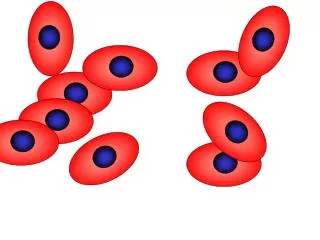
Types of cells in the brain • Neuron: Cell which is responsible for receiving, transmitting and synthesizing information • cell body: contains organelles for metabolism and a nucleus • Glial Cells: Support cells for Neurons (CNS: oligodendrocytes, astrocytes, ependymal cells, radial glial; PNS : Satellite and Schwann cells)
Neuron Types • Neurons come in different types – some only communicate locally, while others have very long axons that communicate with distant regions.
Glial Cells • Glial cells have crucial functions www.mult-sclerosis.org/glialcells.html • Repair, maintenance and cleaning. They produce new myelin when it become damaged, lay down scar tissue, and remove dead cells and other debris. • Physical support. They have hairlike filaments which hold the neurons in place and allow the central nervous system to retain its structural integrity. • CNS development. Help migration of neurons. • Chemical regulation. Supply chemicals such as potassium and calcium and regulate neurotransmitter levels. • Ten times as many glial cells as neurons
Multiple choice • Why is the difference between a tumor and a cancer? • Cancer involves neurons, tumors involve other cells (e.g. glial cells). • Tumor involves neurons, cancer involves other cells (e.g. glial cells). • Tumor is due to virus, cancer is due to bacteria • A tumor can be benign, pre-malignant or malignant, whereas cancer is by definition malignant.
Tumors • Tumor from the Latin "swelling“ • In medicine, swelling due to abnormal, uncontrolled cell division • Brain tumors are usually due to glial cells (gliomas). • Glial cells more common, so higher probability of cell becoming cancerous. • Neuron’s usually stop dividing earlier.
The Central Nervous System • Telencephalon (Cerebrum) • Cortex • Basal Ganglia • Diencephalon • Thalamus • Hypothalamus) • Mesencephalon (Midbrain) • Colliculi • Substancia Nigra • Rhombencephalon • Cerebellum • Pons • Medulla
Deep Structures • Basal Ganglia – Initiates movements • Caudate nucleus, Putamen,Globus pallidus • Diencephalon • Thalamus: Relay from body to cortex • Hypothalamus and pituitary gland: Regulation (e.g. hormone secretion)
Deep Structures • Basal Ganglia – Initiates movements • Caudate nucleus(red) • Putamen (green) • Globus pallidus (blue) • Diencephalon • Thalamus: (yellow) • Hypothalamus: (not shown)
Brain Stem • Midbrain • Early auditory/visual processing • Dopamine for movement control • CN III and IV emerge • Pons • CN V, VI, VII VIII • Medulla Oblongata • Pyramidal decussation: nerves from left cross to right side and vise versa • CN IX, X, XI, XII
The cortex • Cortex – ‘Bark’ shell of brain – mostly gray matter ~80% of human brain ~20% of squirrel brain
Multiple choice • What does ‘temporal’ usually refer to? • Space. • Color. • Time. • Loudness.
Multiple choice • Why is it called the temporal lobe? • This area handles memory – remembering previous times. • This lobe processes hearing – hearing requires good temporal precision. • This lobe is under the temples, where the hair turns gray early in life. • This area helps with counting –which we use for timing events.
Cortical folding • Cortical folding increases surface area. • Ridges are called Gyri (singular = Gyrus) • Greek gyros = circle, hence a coil of brain cortex • Valleys are called Sulci (singular = Sulcus). • Latin = a groove. Gyri Sulci
Gray and White Matter • The outer surface of the cortex is gray matter: lots of interconnected neurons (like cities) • Underneath is the white matter – the highways connecting regions.
Functional Classifications • Some neurons transmit general information • Pain and Temperature • Originate in surface structures • Other neurons transmit specialized information • Specialized receptors • Hearing and vision • Somatic: Skeletal muscles • Visceral: Refer to internal vital body organs • Can be either • Afferent: Sensory • Efferent: Motor
Cortical layers • Neurons are in six layers I. Molecular layer II. External granular layer III. External pyramidal layer IV. Internal granular layer V.Internal pyramidal layer VI. Fusiform layer • Functions • Superficial layers (I-III): inter-cortical connections • IV: input from thalamus • V,VI: outputs to leave cortex