Joints (Articulations)
Joints (Articulations). Case: Hyaluronic acid injections for knee osteoarthritis Joseph, a 45 year old construction worker has osteoarthritis. His knees have been bothering him and his physician recommended injections of hyaluronic acid. Articulation—site where two or more bones meet

Joints (Articulations)
E N D
Presentation Transcript
Joints (Articulations) Case: Hyaluronic acid injections for knee osteoarthritis Joseph, a 45 year old construction worker has osteoarthritis. His knees have been bothering him and his physician recommended injections of hyaluronic acid. • Articulation—site where two or more bones meet • Functions of joints: • Give skeleton mobility • Hold skeleton together
Functional Classification of Joints • Based on amount of movement allowed by the joint • Three functional classifications: • Synarthroses—immovable • Amphiarthroses—slightly movable • Diarthroses—freely movable
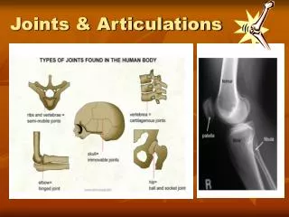
Structural Classification of Joints • Based on material binding bones together and whether or not a joint cavity is present • Three structural classifications: • Fibrous • Cartilaginous • Synovial
Fibrous Joints • Bones joined by dense fibrous connective tissue • No joint cavity • Most are synarthrotic (immovable) • Three types: • Sutures • Syndesmoses • Gomphoses
Fibrous Joints: Sutures • Rigid, interlocking joints containing short connective tissue fibers • Allow for growth during youth • In middle age, sutures ossify and are called synostoses
Crouzon's and Apert's Syndrome Crouzon and Apert syndromes are the most common of the craniosynostosis syndromes. Craniosynostosis refers to the early closing of one or more of the sutures of an infant's head. The skull is normally composed of bones which are separated by sutures. This diagram shows the different sutures which can be involved. As an infant's brain grows, open sutures allow the skull to expand and develop a relatively normal head shape. If one or more of the sutures has closed early, it causes the skull to expand in the direction of the open sutures. This can result in an abnormal head shape. In severe cases, this condition can also cause increased pressure on the growing brain.
(a) Suture Joint held together with very short, interconnecting fibers, and bone edges interlock. Found only in the skull. Suture line Dense fibrous connective tissue Figure 8.1a
Fibrous Joints: Syndesmoses • Bones connected by ligaments (bands of fibrous tissue) • Movement varies from immovable to slightly movable • Examples: • Synarthrotic distal tibiofibular joint • Diarthrotic interosseous connection between radius and ulna
(b) Syndesmosis Joint held together by a ligament. Fibrous tissue can vary in length, but is longer than in sutures. Fibula Tibia Ligament Figure 8.1b
Fibrous Joints: Gomphoses • Peg-in-socket joints of teeth in alveolar sockets • Fibrous connection is the periodontal ligament
(c) Gomphosis “Peg in socket” fibrous joint. Periodontal ligament holds tooth in socket. Socket of alveolar process Root of tooth Periodontal ligament Figure 8.1c
Cartilaginous Joints • Bones united by cartilage • No joint cavity • Two types: • Synchondroses • Symphyses
Cartilaginous Joints: Synchondroses • A bar or plate of hyaline cartilage unites the bones • All are synarthrotic
(a) Synchondroses Bones united by hyaline cartilage Sternum (manubrium) Epiphyseal plate (temporary hyaline cartilage joint) Joint between first rib and sternum (immovable) Figure 8.2a
Asymmetric Closure of Ischiopubic Synchondrosis in Pediatric Patients: Correlation with Foot Dominance OBJECTIVE. The enlarged ischiopubic synchondrosis is a well-known anatomic struc- ture; however, little is known about its physiology. In early childhood, enlargement of this synchondrosis occurs bilaterally, whereas before complete ossification, it is frequently found unilaterally. In most children, the unilateral enlarged ischiopubic synchondrosis is observed in the left hemipelvis, a finding that was hitherto unexplained. During common athletic activi- ties, increased ground reaction forces are exerted on the weight-bearing nondominant limb, which in up to 87% of the general population is the left leg. The asymmetric exertion of these forces may explain the distinct closure sequence of this temporary joint. The purpose of this study was to correlate unilateral enlarged ischiopubic synchondrosis with foot dominance. MATERIALS AND METHODS. The study cohort comprised 32 children who had un- dergone unenhanced radiography, CT, or MRI for reasons other than bone disorders and who presented with enlarged ischiopubic synchondroses. In these children, the distribution of en- larged ischiopubic synchondrosis and foot dominance were evaluated either retrospectively (n = 11) or prospectively (n = 21). RESULTS. In this cohort, 78% of patients were right-footed and 22% were left-footed. Nine of the 32 children presented with unilateral enlarged ischiopubic synchondrosis (left, seven [78%] of nine; right, two [22%] of nine). All children with enlarged left ischiopubic synchondrosis were right-footed, and all children with enlarged right ischiopubic synchon- drosis were left-footed. CONCLUSION. Unilateral enlarged ischiopubic synchondrosis is closely correlated with foot dominance. The asymmetric ossification pattern of the ischiopubic synchondrosis indicates delayed ossification of this anatomic structure due to asymmetrically applied me- chanical forces to the nondominant limb.
Cartilaginous Joints: Symphyses • Hyaline cartilage covers the articulating surfaces and is fused to an intervening pad of fibrocartilage • Strong, flexible amphiarthroses
(b) Symphyses Bones united by fibrocartilage Body of vertebra Fibrocartilaginous intervertebral disc Hyaline cartilage Pubic symphysis Figure 8.2b
Diastasis symphysis pubis is the separation of normally joinedpubic bones, as in the dislocation of the bones, without a fracture.
Synovial Joints • All are diarthrotic • Include all limb joints; most joints of the body
Synovial Joints Distinguishing features: • Articular cartilage: hyaline cartilage • Joint (synovial) cavity: small potential space
Synovial Joints Distinguishing features: 3. Articular (joint) capsule: • Outer fibrous capsule of dense irregular connective tissue • Inner synovial membrane of loose connective tissue
Synovial Joints Distinguishing features: 4. Synovial fluid: • Viscous slippery filtrate of plasma + hyaluronic acid • Lubricates and nourishes articular cartilage Case: Hyaluronan is also used to treat osteoarthritis of the knee. Such treatments, called viscosupplementation, are administered as a course of injections into the knee joint, and are believed to supplement the viscosity of the joint fluid, thereby lubricating the joint, cushioning the joint, and producing an analgesic effect.
Osteoarthritis is caused by 'wear and tear' on a joint. Cartilage is the firm, rubbery tissue that cushions your bones at the joints, and allows bones to glide over one another. Cartilage can break down and wear away. As a result, the bones rub together, causing pain, swelling, and stiffness. Bony spurs or extra bone may form around the joint, and the ligaments and muscles become weaker and stiffer.
Ligament Joint cavity (contains synovial fluid) Articular (hyaline) cartilage Fibrous capsule Articular capsule Synovial membrane Periosteum Figure 8.3
Synovial Joints Distinguishing features: 5. Three possible types of reinforcing ligaments: • Capsular (intrinsic)—part of the fibrous capsule • Extracapsular—outside the capsule • Intracapsular—deep to capsule; covered by synovial membrane
Synovial Joints Distinguishing features: 6. Rich nerve and blood vessel supply: • Nerve fibers detect pain, monitor joint position and stretch • Capillary beds produce filtrate for synovial fluid
The cause of RA is unknown. It is an autoimmune disease, which means the body's immune system mistakenly attacks healthy tissue. RA can occur at any age, but is more common in middle age. Women get RA more often than men. Infection, genes, and hormone changes may be linked to the disease.
Synovial Joints: Friction-Reducing Structures • Bursae: • Flattened, fibrous sacs lined with synovial membranes • Contain synovial fluid • Commonly act as “ball bearings” where ligaments, muscles, skin, tendons, or bones rub together
Coracoacromial ligament Subacromial bursa Humerus resting Cavity in bursa containing synovial fluid Bursa rolls and lessens friction. Humerus head rolls medially as arm abducts. Humerus moving (b) Enlargement of (a), showing how a bursaeliminates friction where a ligament (or otherstructure) would rub against a bone Figure 8.4b
Prepatellar bursitis, also known as Housemaid's knee, is a common cause of swelling and pain above the patella (kneecap), and is due to inflammation of the prepatellar bursa. This structure is a superficial bursa with a thin synovial lining located between the skin and the patella. The bursa develops within the first years of life as a result of mechanical pressure and friction, and it serves the purpose of reducing friction on underlying structures and allowing maximal range of motion in the knee. Aseptic prepatellar bursitis is commonly caused by repetitive work in a kneeling position, hence the name "housemaid's knee"...
Synovial Joints: Friction-Reducing Structures • Tendon sheath: • Elongated bursa that wraps completely around a tendon
Acromion of scapula Coracoacromial ligament Joint cavity containing synovial fluid Subacromial bursa Fibrous articular capsule Hyaline cartilage Tendon sheath Synovial membrane Tendon of long head of biceps brachii muscle Fibrous capsule Humerus (a) Frontal section through the right shoulder joint Figure 8.4a
Stabilizing Factors at Synovial Joints • Shapes of articular surfaces (minor role) • Ligament number and location (limited role) • Muscle tone, which keeps tendons that cross the joint taut • Extremely important in reinforcing shoulder and knee joints and arches of the foot
Synovial Joints: Movement • Muscle attachments across a joint: • Origin—attachment to the immovable bone • Insertion—attachment to the movable bone • Muscle contraction causes the insertion to move toward the origin • Movements occur along transverse, frontal, or sagittal planes
Synovial Joints: Range of Motion • Nonaxial—slipping movements only • Uniaxial—movement in one plane • Biaxial—movement in two planes • Multiaxial—movement in or around all three planes
Summary of Characteristics of Body Joints Consult Table 8.2 for: • Joint names • Articulating bones • Structural classification • Functional classification • Movements allowed
Movements at Synovial Joints • Gliding • Angular movements: • Flexion, extension, hyperextension • Abduction, adduction • Circumduction • Rotation • Medial and lateral rotation
Movements at Synovial Joints 4. Special movements • Supination, pronation • Dorsiflexion, plantar flexion of the foot • Inversion, eversion • Protraction, retraction • Elevation, depression • Opposition
Gliding Movements • One flat bone surface glides or slips over another similar surface • Examples: • Intercarpal joints • Intertarsal joints • Between articular processes of vertebrae
Gliding (a) Gliding movements at the wrist Figure 8.5a
Angular Movements Movements that occur along the sagittal plane: • Flexion—decreases the angle of the joint • Extension— increases the angle of the joint • Hyperextension—excessive extension beyond normal range of motion
Hyperextension Extension Flexion (b) Angular movements: flexion, extension, and hyperextension of the neck Figure 8.5b
Extension Hyperextension Flexion (c) Angular movements: flexion, extension, andhyperextension of the vertebral column Figure 8.5c
Flexion Extension Flexion Extension (d) Angular movements: flexion and extension at theshoulder and knee Figure 8.5d