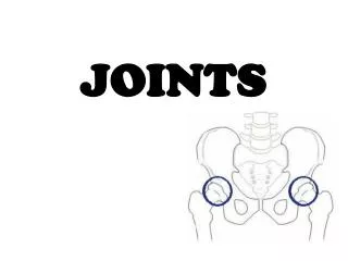
JOINTS
JOINTS. Chapter 9. Introduction . Joints or articulations are sites where two or more bones meet Joints have two fundamental functions: provide for skeletal mobility hold the skeleton together

JOINTS
E N D
Presentation Transcript
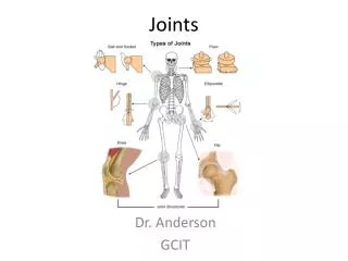
JOINTS Chapter 9
Introduction • Joints or articulations are sites where two or more bones meet • Joints have two fundamental functions: • provide for skeletal mobility • hold the skeleton together • Weakest parts of the skeleton, yet have a remarkable ability to resist the forces that tear them apart
Classification of Joints • Structural classification • focuses on the material binding the bones together and whether or not there is a joint cavity (fibrous, cartilaginous, synovial) • Functional classification • based on the amount of movement allowed at the joint (synarthroses, amphiarthoroses, diarthroses)
Functional Classification • Synarthroses • Immovable joints • Amphiarthroses • Slightly movable joints • Diarthroses • Freely movable joints
Structural Classification • Fibrous • Joined by fibrous tissue • Cartilaginous • Joined by cartilage • Synovial • The bones are joined and surrounded by a joint cavity • Note: • Structural classification is the system used in your text
Summary of Joint Classes • Fibrous joints • Suture • Syndesmoses • Gomphoses • Cartilaginous joints • Synchondroses • Symphyses • Synovial • Gliding, hinge, pivot, condyloid, saddle, and ball and socket
Fibrous Joints In fibrous joints the bones are joined by fibrous tissue; no joint is present. The three types of fibrous joints are. . . • Sutures • Dense fibrous connective tissue • Syndesmosis • A cord or band of connective tissue • Gomphosis • Peg-in-socket arrangement surrounded by fibrous tissue or peridontal ligament
Suture Joint • Occurs only between bones of the skull • Wavy articulating bone edges interlock • Junction is filled by connective tissue • Rigid splices bind bones of the skull together tightly
Syndesmosis • Longer fibrous tissue occurs as a sheet or membrane • Longer fibrous tissue permits the joint to “give” or flex • True movement is not possible
Gomphosis • Fibrous tissue holds teeth in their sockets • Teeth embedded in sockets of bone • Anchored by fibers of periodontal ligament
Cartilaginous Joints In cartilaginous joints, the articulating bones are united by cartilage, there is no joint cavity • Synchondroses • Hyaline cartilage unites the bones • Symphyses • Fibrocartilage unites the bones
Synchondroses • Hyaline cartilage unites the bones • Epiphyseal plates in growing children • Provide for bone growth • When growth ends all synchondroses become immovable Epipyseal Plate
Synchrondroses • Sternocostal joint between the manubrium and rib 1 is a immovable hyaline cartilage joint
Symphyses • Bone surfaces are covered with articular hyaline cartilage which is fused to a pad of fibrocartilage • Fibrocartilage is resilient and acts as a shock absorber and permits limited movement
Synovial Joints • In synovial joints articulating bones are located within a fluid containing joint cavity • Synovial joints permit substantial range of motion • All synovial joints have similar features
Structures of Synovial Joint • Articular cartilage • Hyaline cartilage on opposing bone surfaces • Joint (synovial) cavity • Space filled with fluid • Articular capsule • Capsule to confine fluid • Synovial fluid • Fluid to lubricate joints • Reinforcing ligaments • Maintain joint alignment
Articular Cartilage • Hyaline cartilage covers the bone surfaces • Cartilage absorbs the compression placed on the joint • Cartilage keeps the bone ends from being crushed
Joint (synovial) cavity • Joint spaces are unique to synovial joints • Joint spaces are filled with synovial fluid
Articular capsule • The joint cavity is enclosed by a double layered articular capsule • The external layer is a tough flexible fibrous capsule • The inner synovial membrane
Synovial Fluid • Synovial fluid fills the entire joint • Slippery fluid lubricates joint • Weeping lubrication squeezes synovial fluid into and out of the cartilage nourishing the cells Synovial Fluid
Reinforcing ligaments Extracapsular Ligament • Ligaments reinforce joints • Intrinsic ligaments reinforce capsule • Extracapsular are outside capsule • Intracapsular are inside capsule Intracapsular Ligament
Features of Select Synovial Joints • Certain synovial joints have additional structural features • Fatty pads cushion the knee and hip joints • Fibrocartilage articular discs separates articular surfaces (menisci) • Articular discs improve the fit between the articulating surfaces (knee, jaw)
Bursae and Tendon Sheaths • Bursae and tendon sheaths are closely associated with synovial joints • Essentially sacs of lubricant • Function as “ball bearings” to reduce friction between adjacent structures • Reduces friction during joint activity
Bursae • Bursae are flattened fibrous sacs lined with synovial membrane and containing a thin film of synovial fluid • Common at sites where ligaments, skin, muscles or tendons rub against a bone
Bursae: Anomolies • A bunion is an enlarged bursae at the base of the big toe • False bursae may develop at any site where there is excessive motion • Function similar to a true bursae
Tendon Sheaths Tendon Sheath • An elongated bursa that wraps completely around a tendon subjected to friction • Tendon slides within this lubricated sleeve • Common at sites where the tendon is subject to friction from other tendons or bone features
Retinaculum Retinaculum • Retinaculum function to confine tendons to a specific line of pull • Muscle exerts a force around a skeletal feature • Similar to a pulley or gear changing the angle of force exerted by a machine
Factors Influencing Synovial Joint Stability • The stability of a synovial joint depends on three factors . . . • The nature of the articular surfaces • The number and positioning of the ligaments • The tone and strength of the muscles acting upon the joint
Articular Surfaces • The surfaces determine what movements are possible at a joint, but play a minimal role in joint stability • Many joints have shallow, “misfit” surfaces • Larger surfaces or deeper sockets vastly improve stability • Ball and socket joints are very stable because of their articular surfaces
Articular Surfaces • The knee is a hinge joint by classification • The knee is an example of a joint that allows for “extra” movements • The joint surfaces allow for some anterior - posterior sliding, sliding, as well as a slight amount of rotation at full extension
Ligaments • Ligaments unite the bones of a joint • Ligaments help to direct bone movement and prevent excessive or undesirable motion • As a rule, the more ligaments a joint has the stronger it is • Ligaments can stretch due to undue tension or trauma • Ligaments can stretch only 6% of its length before it snaps
Supporting Ligaments • The supporting ligaments of the elbow allow flexion / extension and restrict movement in any other plane • The Annular ligament allows for rotation of the head of the radius but restricts other movements
Muscle Tone • In most joints the muscles that act upon a joint are the most important stabilizing factor • The tendons of the muscles keep the joint taunt and provide dynamic support • Muscle tone is extremely important in reinforcing the shoulder and knee joint as well as the arches of the foot • The articular capsule and the ligament have extensive sensory nerve endings providing reflexive contraction of supporting muscles
Muscle Tone • The knee is a joint that features movement over stability • The knee is very dependent upon the muscles to provide dynamic stability to the joint while it moves • Note: Rehab
Movements Allowed by Synovial Joints • Nonaxial • Biaxial • Multiaxial
Gliding Movements • Simplest type of joint movement • Bone surface glides or slips over another similar surface • Occur at the intercarpal and intertarsal joints as well as articular processes of vertebrae
Flexion/Extension • Flexion • A bending movement that decreases the angle of the joint • Extension • A movement that increases the angle of the joint
Flexion/Extension/Hyperextension • Flexion • A bending movement that decreases the angle of the joint • Extension • A movement that increases the angle of the joint • Hyperextension • Bending beyond the upright position
Flexion • Flexion • A bending movement that decreases the angle of the joint and brings the two articulating bones closer together • Movement usually occurs in the sagittal plane • Illustrated • Flexion of the arm • Flexion of the leg
Extension • Extension • A movement that increases the angle of the joint that moves the two articulating bones farther apart • Movement within the sagittal plane • Illustrated • Extension of the leg and arm
Dorsiflexion and Plantar Flexion • Dorsiflexion • Lifting the foot so that its superior surface nears the shin • Plantar flexion • Depressing the foot or pointing the toes downward
Ab/Adduction/Circumduction • Abduction • Movement of a limb away from midline or a spreading of the digits of the hand or foot • Adduction • Movement of a limb toward midline or in the case of the digits toward the midline of the hand or foot • Circumduction • Movement of a limb in a circle
Rotation • Rotation is the turning of a bone around its own long axis • Only movement possible between C1 & C2 • Common at the hip and shoulder joints • Medial or lateral is a function of whether rotation results in the anterior surface of the limb moving toward or away from the midline of the body
Types of Synovial Joints • Although all synovial joints have the same features they do not have a common structural plan • Based on the shape of their articular surfaces there are six major categories of synovial joints • Plane, hinge, pivot, condyloid, saddle, and ball and socket
Plane Joint • A plane joint is the only example of a nonaxial joint • Articular surfaces are essentially flat • Allow only short slipping or gliding movements