Download
1 / 39
400 likes | 676 Vues
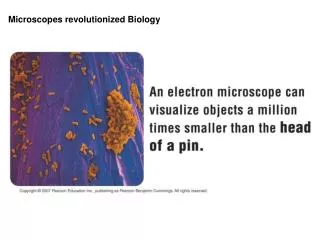
Microscopes revolutionized Biology. Microscopes made cell biology possible. Cell Biology has contributed greatly to advancing medicine and other fields, and led directly to the field of Molecular Biology. With Cells, Size Matters (or the metric system can be fun).

E N D
Cell Biology has contributed greatly to advancing medicine and other fields, and led directly to the field of Molecular Biology.
Mr. Microscope, Antonie van Leeuwenhoek Brass replica showing size . . . my work, which I've done for a long time, was not pursued in order to gain the praise I now enjoy, but chiefly from a craving after knowledge, which I notice resides in me more than in most other men. And therewithal, whenever I found out anything remarkable, I have thought it my duty to put down my discovery on paper, so that all ingenious people might be informed thereof. Antony van Leeuwenhoek. Letter of June 12, 1716 Brass single lens microscope, Univ. of Utrecht
Leeuwenhoek’s work Rotifer Bacteria
Spontaneous Generation -The theory with nine lives Louis Pasteur 1859 John Needham 1745 Lazzaro Spallanzani 1768 Francesco Redi 1668 The controversy over the “spontaneous” generation of life from dead material has an ancient origin. First disproved by Redi for macroscopic life, it became an issue again after the discovery of microbes. Over 100 years later it was finally laid to rest through the work of Spallanzani and the more comprehensive and elegant experiments of Pasteur.
Many different types of light and electron microscopy have been developed. From the top: Euglena as imaged by differential interference contrast light microscopy (uses polarized light to create a pseudo 3D image).Surface detail as revealed by S.E.M. and internal detail as revealed by T.E.M.
Bacterium transmission EM, showing nucleoid region and lack of internal structure
Different plant cells have cell walls of differing thickness. The thicker walls of the vascular bundle fiber and xylem cells (red) have several layers
Plasma membranes are a “fluid mosaic” of proteins in a phospholipid bilayer.
Phospholipids are synthesized by replacing a triglyceride fatty acid with more polar groups
The “Rough” ER creates proteins for export See the following “ribosome” slides for details
The Golgi apparatus and vesicles modify and package materials for export (exocytosis)
Lysosomes are created by the Golgi and contain enzymes that breakdown materials taken into the cell by endocytosis.
Ribosomes consist of two subunits and are the protein manufacturing centers of the cell
Many ribosomes can “read an mRNA blueprint at once. Ribosomes carry out translation of a messenger RNA to make a protein. Small transfer RNAs (trucks) deliver specific amino acids in the right sequence and a protein is made.
The elements of the cytoskeleton are responsible for giving cells shape, allowing them to move, and directing internal organelle “traffic.”
Our ability to manipulate cells in vivo and in vitro is contributing to a medical revolution.