Download
1 / 93
1.11k likes | 1.87k Vues
Clinical Approach to New Onset Arthritis. Jeffrey Carlin, MD Division of Rheumatology, VMMC Clinical Associate Professor, UW. Nothing to declare. Acute Arthritis. The sudden onset of inflammation of the joint, causing severe pain, swelling, and redness.

E N D
Clinical Approach to New Onset Arthritis Jeffrey Carlin, MD Division of Rheumatology, VMMC Clinical Associate Professor, UW
Acute Arthritis • The sudden onset of inflammation of the joint, causing severe pain, swelling, and redness. • Structural changes in the joint itself may result from persistence of this condition.
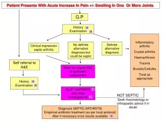
Key Points 1. Distinguish arthritis from soft tissue non- articular syndromes (discrepancy between “active” and “passive” ROM suggests periarticular/soft tissue) 2. If the problem is articular distinguish single joint from multiple joint involvement 3. Inflammatory or non-inflammatory disease 4. Always consider septic arthritis!
Acute Monoarthritis • Inflammation (swelling, tenderness, warmth) in one joint • Occasionally polyarticular diseases can present with monoarticular onset: (RA, JRA,Reactive and enteropathic arthritis, Sarcoid arthritis, Viral arthritis, Psoriatic arthritis)
Acute Monoarthritis - Etiology • THE MOST CRITICAL DIAGNOSIS TO CONSIDER: INFECTION !
Acute Monoarthritis - Etiology • Septic • Crystal deposition (gout, pseudogout) • Traumatic (fracture, internal derangement) • Other (hemarthrosis, osteonecrosis, presentation of polyarticular disorders)
Questions to Ask – History Helps in Differential Diagnosis • Pain come suddenly, minutes? – fracture. • 0ver several hours or 1-2 days? –infectious, crystals, inflammatory arthropathy. • History of IV drug abuse or a recent infection? – septic joint. • Previous similar attacks? – crystals or inflammatory arthritis. • Prolonged courses of steroids? – infection or osteonecrosis of the bone.
Indications for Arthrocentesis • SYNOVIAL FLUID ANALYSIS: The single most useful diagnostic study in initial evaluation of monoarthritis • 1. Suspicion of infection • 2. Suspicion of crystal-induced arthritis • 3. Suspicion of hemarthrosis • 4. Differentiating inflammatory from noninflammatory arthritis
Tests to Perform on Synovial Fluid • Gram stain and cultures • Total leukocyte count/differential • Inflammatory vs. non-inflammatory • Polarized microscopy to look for crystals • Not necessary routinely: • Chemistry (glucose, total protein, LDH) unlikely to yield helpful information beyond the previous tests.
Other Tests Indicated for Acute Arthritis 1. Almost always indicated: Radiographs CBC ESR/CRP 2. Indicated in certain patients: Cultures 3. Rarely indicated: Serologic: ANA, RF, HLA-B27 Serum Uric acid level
Tests of Acute Phase Reactants • Erythrocyte Sedimentation Test • C-Reactive Protein
Patterns of Response of Acute Phase Reactants Gabay C, Kushner I, NEJM , 1999;340:450
ESR’s • Non-specific marker- elevated in rheumatic diseases, infection, malignancy • Can be artificially elevated by: • Pregnancy • Anemia • Nephrotic Syndrome • Benign/Malignant Monoclonal Gammopathies • Age • Obesity • Can be normal in some inflammatory conditions
Formula for Age- Related Normals • Men: ESR(mm/hr)= (age in years)/2 • Females ESR (mm/hr)= (age in years + 10)/2
C- Reactive Protein • Produced in liver in response to IL-1 & IL-6 • Rapid rise in response to inflammatory stimuli • Can be affected by: • Obesity/Metabolic Syndrome • Age
Formula for Age-Related Normals • Men CRP = (age/65) +.1 mg/dl • Women CRP = (age/65) + .7 mg/dl
Septic Joint • Most articular infections – a single joint • 15-20% cases polyarticular • Most common sites: knee, hip, shoulder • 20% patients afebrile • Joint pain is moderate to severe • Joints visibly swollen, warm, often red • Comorbidities: RA, DM, SLE, cancer,etc
Septic Joint - Nongonococcal • 80-90% monoarticular • Most develop from hematogenous spread • Most common: • Gram positive aerobes (80%) • Majority with Staph aureus (60%) • Gram negative 18%
Gout • Caused by monosodium urate crystals • Most common type of inflammatory monoarthritis • Typically: first MTP joint, ankle, midfoot, knee • Pain very severe; cannot stand bed sheet • May be with fever and mimic infection • The cutaneous erythema may extend beyond the joint and resemble bacterial cellulitis
Urate Crystals • Needle-shaped • Strongly negative birefringent
Pseudogout • Can cause monoarthritis clinically indistinguishable from gout. • Often precipitated by illness or surgery. • Pseudogout is most common in the knee (50%) and wrist. • Reported in any joint (Including MTP). • CPPD disease may be asymptomatic (deposition of CPP in cartilage).
CPPD Crystals • Rod or rhomboid-shaped • Weakly positive birefringent
Polyarthritis • Definite inflammation (swelling, tenderness, warmth of > 5 joints • A patient with 2-4 joints is said to have pauci- or oligoarticular arthritis
Acute Polyarthritis Infection • Gonococcal • Meningococcal • Lyme disease • Rheumatic fever • Bacterial endocarditis • Viral (rubella, parvovirus, Hep. B)
Acute Polyarthritis Infection • Gonococcal • Meningococcal • Lyme disease • Rheumatic fever • Bacterial endocarditis • Viral (rubella, parvovirus, Hep. B) Inflammatory • RA • JRA • SLE • Reactive arthritis • Psoriatic arthritis • Polyarticular gout • Sarcoid arthritis
Temporal Patterns in Polyarthritis • Migratory pattern: • Rheumatic fever, gonococcal (disseminated gonococcemia), early phase of Lyme disease • Additive pattern • RA, SLE, psoriasis • Intermittent: • Gout, reactive arthritis
Patterns of Joint Involvement • Symmetric polyarthritisinvolving small and large joints: viral, RA, SLE, one type of psoriatic (the RA-like). • Asymmetric, oligo- and polyarthritisinvolving mainly large joints, preferably lower extremities, especially knee and ankle : reactive arthritis, one type of psoriatic, enteropathic arthritis. • DIP joints: Psoriatic.
Rheumatoid Arthritis • Symmetric, inflammatory polyarthritis, involving large and small joints • Acute, severe onset 10-15 %; subacute 20% • Hand characteristically involved • Acute hand deformity: fusiform swelling of fingers due to synovitis of PIPs • RF/Anti-CCP Ab may be negative at onset and may remain negative in 15-20%! • RA is a clinical diagnosis, no laboratory test is diagnostic, just supportive!
Rheumatoid Factors • Autoantibodies to the Fc portion of IgG. • Support a diagnosis of Rheumatoid Arthritis but are not by themselves diagnostic. • Are seen in about 75% to 80% of patients with RA. • Are associated with a poor prognosis in patients with RA. • Are seen in conditions other than RA
Rheumatic Diseases with Positive RF • RA 80% • JRA 20% • SLE 20% • Sjogren’s 90% • Scleroderma 20-30%
Non-Rheumatic Diseases with Positive RF • Hepatitis C < 70% • Mixed cryoglobulinemia 90% • Sarcoidosis 5-30% • Pulmonary Fibrosis 20% • Infections varies • Aging 5%
RF: Clinical Significance • Highly predictive of RA in patients with identified rheumatic disease • May be absent at the onset of disease in up to half of patients with typical clinical picture of RA • approx 20% remain seronegative • many convert within 2 years • Best used to confirm RA for typical presentation • inflammatory polyarthritis, “gel phenomenon,” etc. • Not useful to follow course of illness • generally not helpful to repeat after diagnosis
RF: Test Statistics • Sensitivity 80% • Specificity 95% • PPV (unselected populations)- 20-30% (RA population)- 80% • NPV- 95%
Anti-Citrulline Antibody Assay ELISA detects antibodies to cyclic citrullinated protein (anti-CCP)
Anti-CCP Antibody Assay • Accuracy (Anti-CCP-2 Assay) • Specificity 79% Sensitivity 96-98% • Diagnosis more accurate when combined with RF+ • Present in 50-60% early RA patients • Can be seen 1.5 -9 yrs pre-diagnosis of RA • Predictive for progressive joint damage • Present in up to 40% percentage of RF- patients with erosions • RF+, anti-CCP+ pts have very aggressive disease
Viral Arthritis • Younger patients • Usually presents with prodrome, rash • History of sick contact • Polyarthritis similar to acute RA • Prognosis good; self-limited • Examples: Parvovirus B-19, Rubella, Hepatitis B and C, Acute HIV infection, Epstein-Barr virus, mumps
Parvovirus B-19 • The virus of “fifth disease”, erythema infectiosum (EI). • Children “slapped cheek”; adults flu-like illness, maculopapular rash on extremities. • Joints involved more in adults (20% of cases). • Frequently RF + • Abrupt onset symmetric polyarthralgia/polyarthritis with stiffness in young women exposed to kids with E.I. • May persist for a few weeks to months.