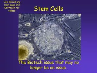
Vascular stem cells and progenitors
Vascular stem cells and progenitors. Lipnik Karoline Department of Vascular Biology and Thrombosis Research Medical University of Vienna Lazarettgasse 19 A-1090 Vienna Austria Basic seminar 1, Vascular Biology, N090/N094, SS 2008 16. June 2008. Overview.

Vascular stem cells and progenitors
E N D
Presentation Transcript
Vascular stem cells and progenitors Lipnik Karoline Department of Vascular Biology and Thrombosis Research Medical University of Vienna Lazarettgasse 19 A-1090 Vienna Austria Basic seminar 1, Vascular Biology, N090/N094, SS 2008 16. June 2008
Overview • Development of blood vessel system – vasculogenesis, angiogenesis • Embryonic stem cells • Alternatives to embryonic stem cells • Adult stem and progenitor cells and prospective therapeutical applications
Vascular development – the beginning cygote blastocyste Germ layers give rise to development of defined cell types http://www.hhmi.org/biointeractive/stemcells/animations.html
ENDODERM Lungs Liver Pancreas Intestine Molecular mechanisms of stem-cell identity and fate Fiona M. Watt and Kevin Eggan
EKTODERM Brain Skin, Hair Mammary gland Molecular mechanisms of stem-cell identity and fate Fiona M. Watt and Kevin Eggan
MESODERM – VASCULAR SYSTEM Haematopoiesis Heart Muscle Mesenchyme Vascular cells Molecular mechanisms of stem-cell identity and fate Fiona M. Watt and Kevin Eggan
Blood vessel formation • Two categories: a.) vasculogenesis: de novo blood vessel generation from vascular progenitor cells b.) angiogenesis: formation of new blood vessels via extension or remodeling of existing blood vessels;
Blood vessel formation • Vasculogenesis: a.) during embryonic development; b.) during adulthood associated with circulating progenitor cells • Angiogenesis: a.) embryonic development b.) adulthood: wound healing, menstrual cycle, tumour-angiogenesis…
Vasculogenesis • The vascular system is one of the earliest organ system that developes during embryogenesis • One of the first markers of angioblast precursors Flk-1 (VEGF-R2) • Further important early markers are: Brachyury and C-Kit
Vasculogenesis 1. First phase • Initiated from the generation of hemangioblasts; leave the primitive streak in the posterior region of the embryo; a part of splanchnic mesoderm 2. Second phase • Angioblasts proliferate and differentiate into endothelial cells 3. Third phase • Endothelial cells form primary capillary plexus
Vasculogenesis • Extraembryonic Vasculogenesis • Intraembryonic Vasculogenesis
Extraembryonic Vasculogenesis • First apparent as blood islands in yolk sac • Blood islands are foci of hemangioblasts • Differentiate in situ: a.) loose inner mass of embryonic hematopoietic precursors b.) outer layer of angioblasts • by the merge of individual blood islands capillary networks are formed • Yolk sac vasculogenesis communicate with fetal circulation via the vitelline vein
Human yolk sac with blood island yolk sac endoderm endothelial cell blood blood island mesoderm Cellular composition of the yolk sac mesoderm Blood island endoderm
Intraembryonic vasculogenesis • para-aortic mesoderm = AGM (aorta-gonad-mesonephros) • First dorsal aorta and cardinal veins are built • Endocardium - vascular plexus is generated • Development of bilateral embryonic aortae • Then allantoic vasculature occurs
Intraembryonic vasculogenesis • Subsequent vascular development primarly via angiogenesis • Some endoderm derived organs, however, are also capable for vasculogenesis
Developmental angiogenesis • Majority of vascular development occurs via angiogenesis • Growth of new blood vessels from existing vessels • Two distinct mechanisms available a.) sprouting angiogenesis b.) intussusceptive angiogenesis
Sprouting angiogenesis • Sprouting: invasion of new capillaries into unvascularized tissue from existing mature vasculature - degradation of matrix proteins - detachment and migration of ECs - proliferation Guided by endothelial tip cells and influenced by various attractant and repulsive factors (Ephrin, Netrin, Plexin…)
Intussusceptive angiogenesis • Intussusceptive or non sprouting angiogenesis: - remodelling of excisting vessels - vessel enlarges - pinches inward - splits into two vessels
Intussusceptive angiogenesis Das Endothel ein multifunktonelles Organ: Entdeckung, Funktionen und molekulare Regulation Stürzl M., et al Cell Tissue Res (2003) 314:107–117 DOI 10.1007/s00441-003-0784-3
Building of blood vessels in adulthood Endothelial precursors Angiogenic sprouting Intussusceptive growth
Important factors guiding angiogenesis • bFGF: proliferation, differentiation, maturation • TGFb: stabilize the mature capillary network by strengthen the ECM structures • PDGF: recruits the pericytes to provide the mechanical flexibility to the capillary • MMP inhibitors: suppresses angiogenesis • Endostatin: cleaved C-terminal fragment of collagen XVII; binds to VEGF to interfere the binding to VEGFR
Summary part 1 • Vascular system developes from mesodermal germ layer • Two categories of vessel building: a.) vasculogenesis: vascular progenitors b.) angiogenesis: sprouting, intussusceptive; from preexisting vessels • Extraembryonic vasculogenesis: yolk sac, blood islands, vascular plexus • Intraembryonic vasculogenesis: AGM region – dorsal aorta and cardinal veins • Majority of blood vessels built by angiogenesis (embryo and adult) • Proangiogenic factors: VEGF, bFGF, angiopoietins • Maturation and stabilization: TGFß and PDGF • Anti-angiogenic: MMP inhibitors, Endostatin
Embryonic stem cells as a tool to study vascular development • generation of stem cells • Differentiation to various cell types http://www.hhmi.org/biointeractive/stemcells/animations.html
Embryonic stem cells • Generation http://www.hhmi.org/biointeractive/stemcells/animations.html
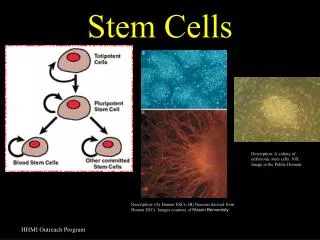
Embryonic stem cells as a tool to study vascular development Characteristics • derived from blastocyst: 3-5 day-old embryo • Unspecialized - totipotent • potential to develop all different cell types • Divide without limit: long term self renewal Tests to identify embryonic stem cells • Subculturing for many months • Specific surface markers: Oct-4, Sox2, NANOG • Testing if cells are pluripotent: differentiation in cell culture; • injecting in vivo - teratoma should be built
Differentiation of ES cells to vascular cells • Stem cells cultivated with a defined cocktail mix (BMP-4, VEGF, SCF, Tpo, Flt3-ligand) in serum free medium to generate embryoid bodies • EBs dissociated and cultivated in specific medium or EBs seeded for outgrowth
KDRlow/C-Kitneg population gives rise to cardiomyocytes, SMCs and Ecs – common progenitor Lei Yang et al., Nature Letters, 2008
Generation of functional hemangioblasts from embryonic stem cells LDL – red vWF - green LDL – red VE-cad- green CD31– red vWF - green
Summary part 2 • Embryonic stem cells are generated from the inner cell mass of blastocystes (3 – 5 dpc) • Characteristics: indefinite life span, totipotent – can give rise to every cell type • Primarly cultivated on feeder cells for expansion of undifferentiated cells • Generation of ECs through stimulation with various cytokine cocktail – Embryoid bodies, outgrowth
Alternatives to human embryonic stem cells • Stem cells derived from single blastomeres • Stem cells through nuclear reprogramming – overview • Induced pluripotent stem cells (iPS) through expression of stem cell specific proteins in differentiated cells
Human embryonic cell lines derived from single blastomeres ectoderm 3-tubulin endoderm alpha-fetoprotein mesoderm SMC Figure 1. Derivation and Characterization of hESC Lines from Single Blastomeres without Embryo Destruction (A) Stages of derivation of hES cells from single blastomere. (a) Blastomere biopsy, (b) biopsied blastomere (arrow) and parent embryo are developing next to each other, (c) initial outgrowth of single blastomere on MEFs, 6 days, and (d) colony of single blastomere-derived hES cells.
Stem cells through nuclear reprogramming - overview • Adult and stem cells are genetically equivalent • Differential gene expression is a result of epigenetic changes during development • Nuclear reprogramming: reversal of the differentiation state of a mature cell to one that is characteristic of the undifferentiated embryonic state A. Nuclear transfer B. Cellular fusion C. Cell extracts D. Culture induced reprogramming
Stem cells through nuclear reprogramming - overview Nuclear transfer Experimental Approach: Reproductive cloning: functional test for reprogramming to totipotency Somatic cell nuclear transfer: efficient derivation of genetically matched ES cells with normal potency Mechanistic insights: Allows epigenetic changes (reversible) to be distinguished from genetic changes (irreversible) Limitations: Reproductive cloning is very inefficient There are abnormalities at all stages of development
Nuclear exchange to generate stem cells http://www.hhmi.org/biointeractive/stemcells/animations.html
Cybrid embryos - human chromosomes with animal eggs In vitro fertilization Intracytoplasmic sperm injection Somatic cell nuclear transfer
Stem cells through nuclear reprogramming - overview Cell fusion Experimental Approach: Nuclear reprogramming of somatic genome in hybrids generated with pluripotent cells; in most hybrids less differentiated partner is predominant Mechanistic insights: Allows study of genetics of reprogramming Question: chromosomes of somatic cells reprogrammed or silenced; nucleus or cytoblast required for molecular reprogramming Limitations: Fusion rate is very low Tetraploid cells are generated
Stem cells through nuclear reprogramming - overview Cell extract Experimental Approach: Exposure of somatic nuclei or permeabilized cells to extracts from oocytes or pluripotent cells; Mechanistic insights: Allows biochemical and kinetic analysis of reprogramming Limitations: No functional reprogramming done
Stem cells through nuclear reprogramming - overview Cell explantation Experimental Approach: Explantation in culture selects for pluripotent, reprogrammed cells; certain physiological conditions entire cells can de-differentiate (Teratokarzinoma) Mechanistic insights: Allows study of genetics of reprogramming Limitations: May be limited to germ line cells
Stem cells through nuclear reprogramming - overview • Molecular mediators of reprogramming and pluripotency • Chromatin remodelling factors • DNA modification • Histone modification • Pluripotency maintained by a combination of extra- and intracellular signals • Extracellular signals: STAT3, BMP, WNT • Intracellular signals: factors at transcriptional level (Oct-4, Nanog, Sox2…)
Stem cells through nuclear reprogramming - overview • Downstream targets: transcription factors, which are silent in undifferentiated cells • Polycomb group (PcG) proteins – chromatin modifiers – repress developmental pathways • Chromatin formation of many key developmental genes: bivalent domains • Activating and inhibitory marks • Bivalent domains are lost in differentiated cells
Induced pluripotent stem cells (iPS) and cellular alchemy • introducing of factors in fibroblasts - induced pluripotent stem cells • Able to produce many cell types • Initially 24 genes selected • Transduced into mouse embryonic fibroblasts • Resistance gene for G418 under control of Fbx15 promoter, which is only active in pluripotent cells • Drug resistant colonies appeared, which resembled ES cells • Expressed transcripts and proteins considered to be part of ES cell signature • Termed: induced pluripotent stem cells (iPS) • Formed all three germ layers in vitro and in vivo • Best combination: Oct-4, Sox2. c-Myc, Klf4
Summary part 3 • Generation of ESCs from single blastomere • Reprogramming of differentiated cells via: - nuclear transfer: molecular cloning - transduction with stem cell genes
Adult progenitor and stem cells and potential clinical application • Undifferentiated cells found among differentiated ones • Identified in various tissues • mainly generate cell types of the tissue in which they reside • can renew themselves (20 to 30 PD) • Can differentiate • Task: Maintenance and repair