Download

1 / 59
640 likes | 982 Vues
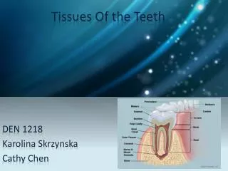
Tissue of the teeth. Dr Jamal Naim PhD in Orthodontics. Enamel. Amelogenesis. Enamel formation Enamel maturation Maturation means also mineralization and does not wait the complete formation of Enamel. Amelogenesis. Enamel formation: Dentino-enamel membrane:

E N D
Tissue of the teeth Dr Jamal Naim PhD in Orthodontics Enamel
Amelogenesis • Enamel formation • Enamel maturation Maturation means also mineralization and does not wait the complete formation of Enamel.
Amelogenesis Enamel formation: • Dentino-enamel membrane: • Induction after dentin formation (first layer is called mantel dentin) • Enamel proteins formed in RER and transported to the distal end with secrete granules and released extracellulary • This is the aprismatic Enamel (dentino-enamel membrane)
Amelogenesis Enamel formation: • Tomes process: • After formation of the DEM, the ameloblasts will move away from dentin and develop the TP. • TP will secrete the granules perpendicular to the membrane of the TP • This direction is responsible for the appearance of the enamel prismatic and inter-prismatic substance.
Amelogenesis Tomes process Rod enamel Tomes process Interrod enamel Rod sheath
Amelogenesis Enamel maturation: • Initial or partial mineralization (immediate) during matrix formation, 25-30% of the total mineralization. • In a second stage the mineralization will be completed, about 96% inorganic substance.
Amelogenesis Enamel maturation: • From cusp tip or incisal edge and progress cervically • The maturation of the crystals begins at its dentinal end and progress to the outer surface. • At first parallel to D.E.J and later to the outer surface of enamel • Maturation occurs by the growth of the primary crystals till they fuse together • The fibrils between the crystals will become thinner.
Maturation occurs by the growth of the primary crystals till they fuse together
Amelogenesis • After eruption, the maturation continues by deposition of ions from saliva to reach 96 % of its weight inorganic substance
COLOUR THICKNESS Physical properties of Enamel PERMEABILITY HARDNESS BRITTLNESS
COLOUR Yellowish white to grayish white; it depends on: • Translucency • Degree of calcification • Homogeneity So: • Yellowishenamel is more translucent, better calcified and homogenous. • Grayish enamel is opaque, less calcified and less homogenous
THICKNESS It is thick at the incisal edge and cusp tip of molars and premolars (2-2.5mm) and ends cervically as knife edge.
Hardness and Brittleness It is the hardest calcified tissue in human body because of its high calcification and crystal orientation. It is greater at the outer surface and decrease at the DEJ. It is greater at the cusp tip or incisal edge and decrease at the cervical line. Although of its hardness, enamel is brittle especially when looses the underlying elastic healthy dentin.
Permeability Enamel acts as a semi-permeable membrane for certain ions from: • the saliva to the outer layer of enamel • the pulp to the inner layer of enamel across dentin.
Histological Structure Ground section Decalcified section the organic substance is burnt and the inorganic substance remain the inorganic substance is dissolved and the organic substance remain
Histological Structure Enamel is formed of: • Enamel crystallites • Enamel Rod • Rod Sheath (packaging of rods) and • Interrod Substance
Histological Structure Enamel crystallites • crystallites are the smallest units of enamel • They are hexagonal in form • about 160 nm in length and 40-70 nm in width
Histological Structure Rod and interrod enamel: • The Tomes’ processorganizes enamel crystallites into rod enamel (prism = about 100 crystallites) and interrod enamel. • Enamel crystallites that elongate near the tip of the Tomes’ process form the rod enamel. • Crystallites that lengthen near the intercellular junctions form the interrod enamel. • The rod enamel and interrod enamel differ in the orientation of their crystallites
Histological Structure Tomes process Rod enamel Tomes process Interrod enamel Rod sheath
Histological Structure The border between rod and interrod enamel is distinct because part of the ameloblast membrane is “nonsecretory,” which creates gaps in the mineralization front. The apatite crystals are oriented parallel to the long axes of the rod in its body and deviate about 65 as they fan out into the margin and the tail.
Histological Structure The rod and interrod enamel differ in the orientation of their crystallites
The enamel rods are arranged in rows with alternating orientation. The interrod enamel
Histological Structure By electron microscopy a common key-hole or paddle-shaperod is seen in cross section. interrod enamel rod enamel
Histological Structure The head of the rod is toward the occlusal or incisal surface where the tail is cervically. coronal cervical
Histological Structure • The number of the enamel rods varies from 5 millions in lower lateral incisor to 12 millions in the upper first permanent molar. • The number of the rods equals the number of the ameloblasts. • At the tooth surface there are about 20000-30000 enamel rods in 1 mm2 • The density of the rods is at the DEJ about 10% higher than at the enamel surface.
Histological Structure • The diameter of the enamel rod is about 5 µm. In key-hole type the height is about 9 µm. • It increases from the dentino-enamel junction to the outer enamel surface by a ratio of 1:2.
Histological Structure The enamel rod is perpendicular to the dentin surface; In deciduous teeth ; the enamel rod is vertical at the cusp tip or incisal edge then become oblique toward the occlusal surface at the middle part and become horizontal at the cervical area (so enamel ends cervically abruptly).
Histological Structure In permanent teeth; the direction of the enamel rods are similar to that of the deciduous teeth at the occlusal 2/3 but at the cervical region are directed root wise (so the enamel ends cervically as a knife edge). permanent teeth deciduous teeth
Histological Structure Course: the enamel rod starts straight at dentino-enamel junction (D.E.J.) for about 30 µ then has a wavy course till near the outer surface of enamel where it become straight once more. Wavy course of enamel rods
Histological Structure Wavy course of enamel rods
Enamel Histology At the incisal edge or cusp tip the enamel rod has a twisted course and is called gnarled enamel Twisted course of enamel rods Gnarled enamel D
Histological Structure Dentino-enamel-Junction • In ground section D.E.J. appears as scalloped line. • The presence of this irregular surface assures the union between enamel and dentine.
Histological Structure Dentin Enamel Dentino-enamel-Junction
Electron micrographs of the Dentino-enamel-Junction The surface of Dentin after removing of Enamel
Histological Structure Enamel spindle: It is an odontoblastic process which extends in between the cells of inner dental epithelium before the formation of enamel.
Histological Structure Enamel Tufts: • They arise from D.E.J. to about 1/5 to 1/3 of the enamel thickness as tufts of grass. • It always appears in transverse ground section. • They are hypo-calcified prisms and inter-prismatic substance. • It takes this shape because of the wavy course of the enamel rod for several layers leading to this tuft form.
Histological Structure Enamel Tufts
Histological Structure • If a tuft runs till the outer enamel layers (more than 1/3 of enamel thickness), then we speak about Enamel lamella (true lamella, Type A lamella). • They are also hypo-calcified prisms and inter-prismatic substance. • True lamella should be distinguished from other types of lamellae (cracks, type B and type C lamella) Enamel lamella
Histological Structure Enamel lamella type A, True lamella Enamel lamella type B Enamel lamella type C hypo-calcified prisms and inter-prismatic substance Enamel cracks post-eruptive, can reach dentine Enamel cracks pre-eruptive, can reach dentine Filled with epithelial cell or connective tissue Filled with organic contentsof saliva It is limited to enamel
Histological Structure To differentiate between true lamella and crack we do careful decalcification: the true lamella will remain where cracks will disappear. Enamel lamella could act as caries spread way.
Histological Structure • During the secretory stage, enamel crystals do not grow continuously, but rather extend in increments. • The enamel rod is formed in a rhythmic manner, every segment of 4 µm in length and formed in a day. It is manifested structurally as prism cross-striations
Histological Structure cross striations 4 µ/day
Histological Structure • More prominent cross-striations occur in a regular period of about every 4 days/1 week and are known as striae of Retzius or incremental lines. • In longitudinal ground section they appear as dark bands reflecting the rhythmic enamel formation. • At the incisal edge and cusp tip they arise from the D.E.J. then go upward and outward surrounding the tip of dentine and come to D.E.J. again so, they do not reach the outer surface of enamel.
Histological Structure striae of Retzius perikymata
Histological Structure striae of Retzius
Histological Structure striae of Retzius
Histological Structure striae of Retzius