Download
1 / 59
590 likes | 623 Vues
Learn about female external genital organs like mons veneris, labia majora, labia minora, clitoris, and internal structures such as the vagina and uterus. Explore functions, relations, and anatomy with detailed information.

E N D
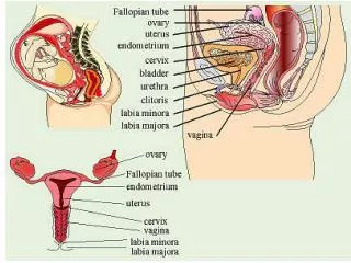
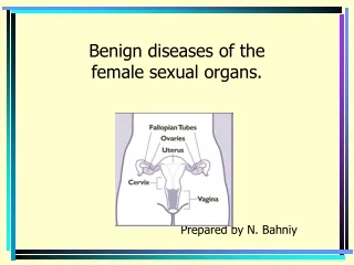
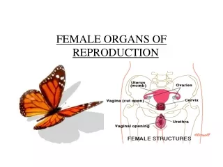
External Genital Organs • The mons veneris/mons pubis • The labia majora • The labia minora • The clitoris • The vestibule • The urethral orifice • The vaginal orifice
This is a pad of fat lying over the symphysis pubis. • It is covered with pubic hair from the time of puberty.
These are two folds of fat and areolar tissue covered with skin and pubic hair on the outer surface.
These are two thin folds of skin lying between the labia majora.
This is a small rudimentary organ corresponding to the male pelvis. • It is extremely sensitive and highly vascular and plays a part in the orgasm of sexual intercourse.
The vestibule • This is the area enclosed by the labia minora in which are situated the openings of the urethra and the vagina.
The urethral orifice • This orifice lies 2.5 cm posterior to the clitoris. The vaginal orifice • This is also known as the introitus of the vagina. • The orifice is partially closed by the hymen,a thin membrane that tears during sexual intercourse or during the birth of the first child.
Bartholin’s glands • These are two small glands that open on either side of the vaginal orifice. • They secrete mucus, which lubricates the vaginal opening.
The vulval blood supply comes from • the internal pudendal arteries • the external pudendal arteries
Lymphatic drainage • This is mainly via the inguinal glands.
Nerve supply • This is derived from the branches of the pudendal nerve.
The vagina • Functions: • The vagina is a passage that allows the escape of the menstrual flow. • Receives the penis and the ejected sperms during sexual intercourse. • Provides an exit for the fetus during delivery.
Relations • Anterior: Bladder and urethra • Posterior: Pouch of Douglas, the rectum and the perneal body. • Superior: Uterus • Inferior: External genitalia
Structure • The posterior wall is 10 cm long • Anterior wall is 7.5 cm in length • The upper end of the vagina is known as the vault • The vaginal walls are pink in appearance and thrown into small folds known as rugae. • These allow the vaginal walls to stretch during intercourse and childbirth.
Contents • There are no glands in the vagina • It is moistened by mucus from the cervix and a transudate that seeps out from the blood vessels of the vaginal wall. • The vaginal fluid is strongly acid ( pH 4.5) owing to the presence of lactic acid formed by the action of doderlein’s bacilli. • These lactobacilli are normal inhabitants of the vagina.
Blood Supply • Branches of the internal iliac artery and includes vaginal artery and a descending branch of the uterine artery.
Lymphatic Drainage • Via the inguinal, the internal iliac and the sacral glands.
Nerve supply • This is derived from the pelvic plexus. • The vaginal nerves follow the vaginal arteries to supply the vaginal walls and also the erectile tissues of the vulva
Functions • The uterus exists to shelter the fetus during pregnancy. • It prepares for this possibility each month and following pregnancy it expels the uterine contents.
Position • The uterus is situated in the cavity of the true pelvis, behind the bladder and in front of the rectum. • It leans forward which is known as anteversion • It bends forwards on itself which is known as anteflexion.
Relations • Anterior: uterovesical pouch and the bladder • Posterior: rectouterine pouch of Douglas and the rectum • Lateral: On either side of the uterus are the broad ligaments, the uterine tubes and the ovaries • Superior: lies the intestines • Inferior: below the uterus is the vagina
Structure • The non pregnant uterus is a hollow ,muscular pear shaped organ situated in the true pelvis • It is 7.5 cm long,5 cm wide and 2.5 cm in depth, each wall being 1.25 cm thick. • The cervix forms the lower third of the uterus and measures 2.5 cm in each direction.
Parts • The body or corpus: This makes up upper two thirds of the uterus and is the greater part. • The fundus: This is a domed upper wall between the insertions of the uterine tubes. • The Cornua: These are the upper outer angles of the uterus where the uterine tubes join. • The cavity: This is a potential space between the anterior and the posterior walls. It is triangular in shape.
The isthmus: This is the narrow area between the cavity and the cervix. It is 7 mm long. • The cervix or the neck: This protrudes into the vagina.The upper half being above the vagina,is known as supra vaginal region. while the lower half is the infra vaginal region.
The internal os ( mouth): This is the narrow opening between the isthmus and the cervix • The external os: This is a small round opening at the lower end of the cervix
Layers • The endometrium: This layer forms a lining of ciliated epithelium on a base of connective tissue or stroma • The myometrium: musle coat: this layer is thick in the upper part of the uterus and is more sparse in the isthmus and cervix • The perimetrium: This is a double serous membrane, an extension of the peritoneum.
Blood supply • Uterine artery • Ovarian artery
Nerve supply • This is mainly from the autonomic nervous system, via the pelvic plexus.
Functions: • The uterine tubes propels the ovum towards the uterus ,receives the sperm as they travel upwards and provides a site for fertilization. • It supplies the fertilized ovum with nutrition during its continued journey to the uterus.
Structure • Each tube is 10 cm long. • Has four portions • The interstitial portion • The isthmus • The ampulla • The infundibulum
The interstitial portion: This is 1.25 cm long and lies within the wall of the uterus • The isthmus: This is another narrow part that extends for 2.5 cm from the uterus • The ampulla: This is a wider portion where fertilization usually occurs. It is 5 cm long • The infundibulum: This is a funnel shaped fringed end that is composed of many processes known as fimbriae.One fimbriae is elongated to form the ovarian fimbria,which is attached to the ovary
Blood supply • Uterine and the ovarian arteries Nerve supply: • This is from the ovarian plexus
Functions • The ovaries produce ova and the hormones oestrogen and progesterone. Positions: The ovaries are attached to the back of the broad ligaments within the peritoneal cavity.
Structure • The medulla • The cortex
The medulla: • This is a supporting framework which is made of fibrous tissue ,the ovarian blood vessels,lymphatics and nerves travel through it. • The cortex • Functioning part of the ovary. • This contains the ovarian follicles in different stages of development.