
SlideServe
Menu
Préférences
© SlideServe · v2026

Clinico-pathologic Findings and Correlations in Anogenital Bowen Disease

vance-pace avatar
4 diapositives · 223 Views · Oct 26, 2014

Clinico-pathologic Findings and Correlations in Anogenital Bowen Disease. Irina Tudose1, M adalina Geanta2, Sabina Zurac3, Florica Staniceanu3, Simona Roxana Georgescu2, V Benea2 1 The Pathology Department, “Prof. Scarlat Longhin” Clinical Hospital
Clinico-pathologic Findings and Correlations in Anogenital Bowen Disease - slide 1
01 / 4Clinico-pathologic Findings and Correlations in Anogenital Bowen Disease
1 / 17

Télécharger la présentation

Clinico-pathologic Findings and Correlations in Anogenital Bowen Disease
Une image / un lien ci-dessous est fourni (tel quel) pour télécharger la présentation

Politique de téléchargement : le contenu du site vous est fourni TEL QUEL pour votre information et votre usage personnel ; il ne peut être vendu, concédé sous licence ni partagé sur d’autres sites sans l’accord de son auteur.Le contenu vous est fourni TEL QUEL, pour votre information et votre usage personnel uniquement. Téléchargez la présentation en cliquant sur ce lien. Lors du téléchargement, si vous ne parvenez pas à télécharger une présentation, l’éditeur a peut-être supprimé le fichier de son serveur.Pendant le téléchargement, si vous ne parvenez pas à obtenir une présentation, le fichier a peut-être été supprimé par l’éditeur.

F I N

Plus de contenu associé

Transcription de la présentation
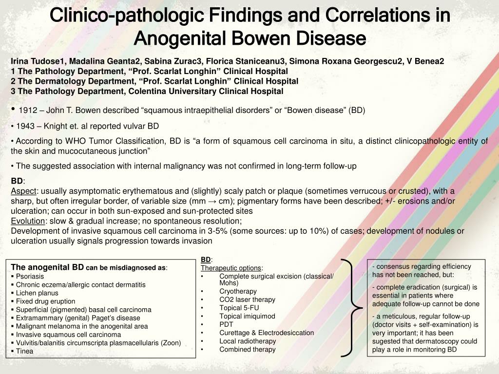
  1. Clinico-pathologic Findings and Correlations in Anogenital Bowen Disease Irina Tudose1, Madalina Geanta2, Sabina Zurac3, Florica Staniceanu3, Simona Roxana Georgescu2, V Benea2 1 The Pathology Department, “Prof. Scarlat Longhin” Clinical Hospital 2 The Dermatology Department, “Prof. Scarlat Longhin” Clinical Hospital 3 The Pathology Department, Colentina Universitary Clinical Hospital • 1912 – John T. Bowen described “squamous intraepithelial disorders” or “Bowen disease” (BD) • 1943 – Knight et. al reported vulvar BD • According to WHO Tumor Classification, BD is “a form of squamous cell carcinoma in situ, a distinct clinicopathologic entity of the skin and mucocutaneous junction” • The suggested association with internal malignancy was not confirmed in long-term follow-up BD: Aspect: usually asymptomatic erythematous and (slightly) scaly patch or plaque (sometimes verrucous or crusted), with a sharp, but often irregular border, of variable size (mm → cm); pigmentary forms have been described; +/- erosions and/or ulceration; can occur in both sun-exposed and sun-protected sites Evolution: slow & gradual increase; no spontaneous resolution; Development of invasive squamous cell carcinoma in 3-5% (some sources: up to 10%) of cases; development of nodules or ulceration usually signals progression towards invasion • BD: • Therapeutic options: • Complete surgical excision (classical/ Mohs) • Cryotherapy • CO2 laser therapy • Topical 5-FU • Topical imiquimod • PDT • Curettage & Electrodesiccation • Local radiotherapy • Combined therapy • The anogenital BD can be misdiagnosed as: • Psoriasis • Chronic eczema/allergic contact dermatitis • Lichen planus • Fixed drug eruption • Superficial (pigmented) basal cell carcinoma • Extramammary (genital) Paget’s disease • Malignant melanoma in the anogenital area • Invasive squamous cell carcinoma • Vulvitis/balanitis circumscripta plasmacellularis (Zoon) • Tinea • consensus regarding efficiency has not been reached, but: • complete eradication (surgical) is essential in patients where adequate follow-up cannot be done • a meticulous, regular follow-up (doctor visits + self-examination) is very important; it has been sugested that dermatoscopy could play a role in monitoring BD

  2. Study: materials and methods • retrospective analysis • we selected 11 patients with histopathological confirmed ano-genital BD and 20 patients with cutaneous BD • all the biopsies were fixed in formaldehyde solution and embedded in paraffin; all the paraffin sections were stained with H-E; immunohistochemestry markers (Ki67, p16, p21, p53) were used for the ano-genital cases • mean age for ano-genital BD was 60,09 years (range 44-74) Most of the patients with ano-genital BD pertained to the age group 60-69, consistent with the literature data (“commonly affects patients in the 6-8th decade”) • The presence of ulceration was evident in 64% of the cases Ano-genital BD cases • In 27% of cases only the labia major was involved, and the rest was equally divided between the labia minor, labia minor+labia major, the frenulum, the perineal region or “the genital region”

  3. The treatment of ano-genital BD cases consisted of: • Complete initial surgical excision (in 3 cases) • Biopsy and HP confirmation, followed by complete surgical excision or curettage and electrodesiccation were surgery was not an option The treatment of extragenital BD cases consisted of: Complete initial surgical excision (in 10 cases) Biopsy and HP confirmation, followed by complete surgical excision in the other 10 cases • Extra-anogenital BD Cases: • Our retrospective study included 20 cases of BD with extragenital location, during last year, with the following features: • mean age 73.8 years (range 55-91); sex ratio 1:1 • most frequent location: the face (40%) • The clinical diagnoses which accompanied the biopsy specimens were: • Bowen’s disease (in 8 cases; the concordance between the clinical and pathological dgn was of 72.72%) • Lichen sclerosus et atrophicus • Genital wart • Erosive genital lichen planus - The concordance between the clinical and pathological diagnosis was of 30%, smaller compared to the situation observed for ano-genital BD - The most frequent clinical confounder was BCC (in 55% of cases)

  4. p53 p16 Ki67 p21 • Histopathological aspects for both ano-genital and cutaneous BD were similar with some particularities regarding ano-genital cases: almost all the lesions were ulcerated, the inflammation was higher consisting in lymphocytes and plasma cells, there were more dyskeratotic cells and a high mitotic rate • Immunohistochemestry analysis for the ano-genital BD cases revealed a high Ki67 value (from 25% to 50%), predominant the lower part of the epithelium, apparently related with p21 high values (from 10% to 60%); p16 presented high positivity (over 20%) in only 3 cases; p53 showed over 10% positivity in 3 cases (8 cases presented less than 5% positivity) • Apparently there is no connection between immunohistochemistry markers values, except for the Ki67 and p21, both with similar positivity in 7 cases of ano-genital BD • The concordance between the clinical and pathological diagnosis was of 30% for the extra-ano-genital cases, smaller compared to the situation observed for ano-genital BD • Having in mind the fact that the observation period was only one year and all the data were collected in a hospital dedicated almost exclusively to dermatology, 11 patients is not such a small number for ano-genital location of BD