Tissue Characterization by Image Analysis for Diagnostic Purposes
200 likes | 338 Vues
This study investigates the characterization of tissue through image analysis, focusing on overcoming various noise types in biomedical imaging modalities. Using ultrasound images, often compromised by speckle noise, the research aims to decompose images into textural and morphological components, associating texture characteristics with diseases such as liver steatosis and atherosclerotic plaques. By advancing classification and quantification techniques, this work seeks to improve diagnostic accuracy and severity assessment through innovative methods in image denoising and analysis.

Tissue Characterization by Image Analysis for Diagnostic Purposes
E N D
Presentation Transcript
Tissue Characterization by Image Analysis for Diagnostic Purposes João Sanches jmrs@ist.utl.pt Institute for Systems and Robotics Instituto Superior Técnico
Noise • Different Biomedical Image Modalities present different types of noise, e.g., additive or multiplicative. • Usually, the noise should be removed. MRI LSFCM US CT
Speckle • Ultrasound images usually present low quality (low SNR) • Images are corrupted by speckle noise (multiplicative) Processed by José Seabra (Biomedical PhD student)
Medical Information However, the speckle pattern contains relevant medical information, e.g., fatty Liver.
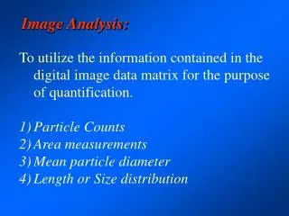
Goal • Decompose the image in textural and anatomical/morphological components. • Characterize the texture/“noise” (speckle) • Associate the texture characteristics with the disease. • Classification and Quantification to Detect (Diagnosis) and Quantify (Severity Assessment) the disease.
Decomposition Noisy Synthetic Image Noiseless Image Noise Field
Ultrasound Image Denoising Noise estimation Intensity and anatomical features Textural features Tissue characterization and Diagnosis Classification
“Noise” Analysis for Diagnostic Purposes Two Cases • Liver Steatosis • Atherosclerotic Plaques (carotids and coronaries)
Steatosis • Steatosis is mainly a textural abnormality of the hepatic parenchyma due to fat accumulation on the hepatic vesicles (genetic, alcohol and obesity) • Today, the assessment is subjectively performed by visual inspection
Steatosis • Comparison with histological data
Steatosis Characterization • Intensity Decay (m) • Texture (Ev,Eh)
Diagnosis Processed by Ricardo Ribeiro (Biomedical PhD student)
3D Diagnosis Global and local analysis
IVUS Collaboration with the Centre de Visió per Computador, Universitat Autònoma de Barcelona, Profª Petia Radeva
IVUS IVUS Image decomposition
IVUS Automatic classification GT
Present and Future Work • New texture characterization and classification methods • Aterosclerotic plaques: 3D Extension of the IVUS • Liver steatosis quantification with additional information from laboratorial analysis data.