Cardiac Cycle
Cardiac Cycle. Ellen Rasche Darcy Holzum Ariel Dunteman. Diastole and Systole . Diastole - Relaxation of the heart Systole - Contraction of the heart. Heart Sounds. Action of the heart valves “lubb” AV valves close and semilunars open Marks the start of the ventricular systole

Cardiac Cycle
E N D
Presentation Transcript
Cardiac Cycle Ellen Rasche Darcy Holzum Ariel Dunteman
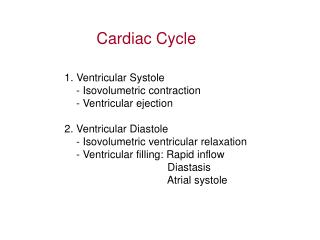
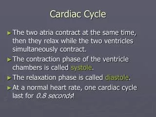
Diastole and Systole • Diastole- Relaxation of the heart • Systole- Contraction of the heart
Heart Sounds • Action of the heart valves • “lubb” • AV valves close and semilunars open • Marks the start of the ventricular systole • Longer than second sound • “dubb” • Beginning of ventricular diastole, when the smilunanr valves close
Heart Sounds (cont.) • Third and fourth sounds may be audible • Usually faint and seldom detectable • Are associated with atrial contraction and blood flowing into the ventricles rather than with valve action
Heart Dynamics • Heart dynamics – refers to movements and forces generated during cardiac contractions • Each time t he heart beats, 2 ventricles eject equal amounts of blood • The stroke volume is the amount ejected by a ventricle during a single beat • Stroke volume varies beat to beat so physicians are often more interested in cardiac output
Heart Dynamics • Cardiac output is the amount of blood pumped by each ventricle in 1 minute • Provides indication of blood flow through peripheral tissues; without adequate blood flow, homeostasis cannot be maintained
Calculate Your Cardiac Output Cardiac output = stroke volume x Heart Rate (mL/min) (mL/beat) (beats/min)
Factors controlling Cardiac Output • The major factors that regulate cardiac output often affect both heart rate and stroke volume • Primary factors include: • Blood volume reflexes • Autonomic innervation • hormones
Blood Volume Reflexes • There is a direct relation between the amount of blood entering the heart and the amount of blood ejected during the contraction • 2 heart reflexes respond to changes in blood volume • Occurs in right atrium and affects heart rate • Ventricular reflex than affects stroke volume
Blood Volume Reflexes con’t • Ariel reflex produces adjustments in heart rate in response to an increase in the venous return • Venous return: flow of venous blood to the heart • The entry of blood stimulates stretch receptors on the right atrial walls, triggering a reflexive increase in heart rate through increased sympathetic activity
Blood Volume Reflexes con’t • The amount of blood pumped out of a ventricle each heartbeat (stroke volume) depends not only venous return but also on the filling time - the duration of ventricular diastole, when blood can flow into the ventricles • The filling time depends primarily on the heart rate: the faster the heart rate, the shorter the available filling time
Blood Volume Reflexes • Over the range of normal activities, the greater the volume of blood entering the ventricles, the more powerful the contraction (and the more pumping force is produced), and the greater the stroke volume • If more blood flows into the heart, the ventricles produce greater force upon contraction • “more in = more out” • This is the Frank-Starling Principle • Output of blood from both ventricles is balanced under a variety of conditions
Factors Controlling Cardiac Output • Autonomic Innervation • The basic heart rate is established by pacemaker cells of the SA node • This rate can be modified by the autonomic nervous system (ANS) • Both they sympathetic and parasympathetic divisions of the ANS innervate the heart • Postganglionic sympathetic fibers extend from neurons • Located in the cervical and upper thoracic ganglia
Autonomic Innervation (cont.) • The vagus nerve carries parasympathetic preganglionic fibers to small ganglia near the heart • Both ANS divisions innervated the SA and AV nodes • Atrial and ventricular cardiac muscles cells
Autonomic Effects on Heart Rate • Reflect the responses of the SA node to acetylcholine (Ach) and to norepinephine (NE) • ACh results in a lower heart rate • Norepinephrine released by sympathetic neurons increases the heart rate • Release of epinephrine € and norepinephrine by the adrenal medullae produces a more sustained rise in heart rate
Autonomic Effect on Stroke Volume • Release of Ne, E and Ach, the ANS affects stroke volume by altering force of myocardial contractions: • Effects of Ne and E: increase the force and degree of contraction, resulting in an increase of stroke volume