Download

1 / 26
270 likes | 810 Vues
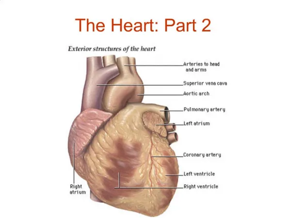
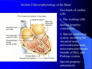
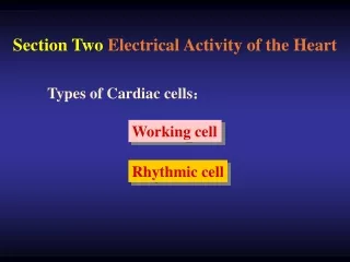
Section 2 Electrophysiology of the Heart. Two kinds of cardiac cells 1, The working cells. Special property: contractility 2, Special conduction system, including the sinoatrial node, atrioventricular node, atrioventricular bundle (bundle of His), and Purkinje system.

E N D
Section 2 Electrophysiology of the Heart Two kinds of cardiac cells 1, The working cells. Special property: contractility 2, Special conduction system, including the sinoatrial node, atrioventricular node, atrioventricular bundle (bundle of His), and Purkinje system. Special property: automaticity
Transmembrane Potentials of Myocardial Cells • 1. Transmembrane Potentials in Working Myocardial Cells • General description • Resting potential: -90mv (-85-95mv) • Action Potential • Phase 0: rapid depolarization, 1-2ms • Phase 1: early rapid repoarization, 10 ms • Phase 2: plateau, slow repolarization, the potential is around 0 mv. 100 – 150ms • Phase 3, late rapid repolarization. 100 – 150 ms • Phase 4 resting potentials 1 2 0 3 4
(2) Ionic bases of transmembrane potentials Resting potential: the equilibration potential of potassium Action potential: Phase 0, Na+ influx. The threshold potential, -70 mv Phase 1, K+ outward flow Phase 2, Ca2+ inward flow and K+ outward flow Phase 3, K+ outward flow Phase 4, Na+ - K+ pump and Na + - Ca2 + exchange (primary and secondary active transport)
2. Transmembrane Potential of Rhythimic Cells • Purkinje cell The phases 0 – 3 are almost the same with that of ventricle cells. At phase 4, the membrane potential does not maintain at a level, but depolarizes automatically – the automaticity Mechanism: If, a kind of Na+ channel that is activated by the hyperpolarization. It is not the same with the fast Na+ channel at phase 0. It could be blocked by Cs but is not affected by TTX .
(2) The SA node cell • Transmembrane potential of the sinoatrial node cell • Maximal repolarization (diastole) potential, –70mv • Low amplitude and long duration of phase 0. It is not so sharp as ventricle cell and Purkinje cell. • No phase 1 and 2 • Comparatively fast spontaneous depolarization at phase 4 A, Cardiac ventricular cell B, Sinoatrial node cell
Mechanism of the membrane potential Phase 0, I Ca-L, slow channel and slow response cell Phase 3, Ik, the time dependent channel, was slowly inactivated near the maximal repolarization potential (-60 mv) Phase 4 Ik, ICa-T and If ICa-T, activated at –50 mv during the repolarization and contribute to the spontaneous deplorization during the Phase 4
Fast and slow response, rhythmic and non-rhythmic cardiac cells • Fast response, non –rhythmic cells: working cells • Fast response, rhythmic cells: cells in special conduction system of A-V bundle and Purkinje network. • Slow response, non-rhythmic cells: cells in nodal area • Slow response rhythmic cells: S-Anode, atrionodal area (AN), nodal –His (NH)cells
II Electrical Properties of Cardiac Cells • Excitability, Conductivity and Automaticity • Excitability of Cardiac Muscle • Factors determining the excitability • Resting potential or maximum diastole potential (rhythmic cell). Low concentration of K+ outside the cell --- resting potential lower – excitability lower • Threshold potential. High concentration of Ca2+ outside the cell – threshold potential less negative – excitability lower • States of Na+ channel. • Resting state: close (could open at the threshold potential); activation (open); inactivation (close, but could not open at any potential), this state will transfer to resting state after a period of repolarization.
(2) Changes in excitability during an action potential • Effective refractory period, including: • A, Absolute refractory period, from the beginning of phase 0 to –60mv of repolarization, no response to stimulus • State of Na+ channel, inactivation • B, Local potential period, form the –60mv to –55mg of phase 3, very strong stimulus can elicit local response but not action potential • State of Na+ channel, most of them are inactivation • Common properties of A and B, from the beginning of phase 0 to –55mv of phase 3, no action potential can be elicited, no matter how strong the stimulus is – effective refractory period • 2) Relative refractory period, from –60 mv to –80 mv of phase 3, a stronger stimulus can elicit action potential, although the duration, amplitude and slope of the upstroke is shorter and smaller • State of Na+ channel, part of them return to the resting state
3) Supernatural period • From –80 mv to –90 mv of phase 3, the excitability is higher than normal • Na+ state. Most of the Na+ channel have returned to the resting condition. • The potential is higher than the resting potential • (3) Relationship between the excitability and contraction of myocardium • No tetanus in cardiac muscle, systole and diastole occur alternately. It is very important for pumping blood to arteries. • Premature excitation, premature contraction and compensatory pause • Extra-stimulus – premature excitation – premature contraction – compensatory pause
2. Automaticity (Autorhythmicity) • Concept: Some tissues or cells have the ability to produce spontaneous rhythmic excitation without external stimulus. • Different intrinsic rhythm of rhythmic cells • Purkinje fiber, 15 – 40 /min • Atrioventricular node 40 – 60 /min • Sinoatrial node 90 – 100 /min • Concept: normal pacemarker, latent pacemarker, ectopic pacemarker • (2) The mechanism that SA node controls the hearts rhythm (acts as pacemaker) rather than the AV node and Purkinje fiber • The capture effect • Overdrive suppression • (3) Factors determining automaticity
Depolarization rate of phase 4 • 2)Threshold potential • 3)The maximal repolarization potential
3. Conductivity • Pathways and characteristics of conduction in heart • Pathways: S-A node -- A-V node --- Bundle of His --- R.L. bundle branches --- Purkinje network – ventricular muscles • Conductive speed of different cardiac muscles: • Atrial myocardium, 0.4m/s; nodal area of A-V junction, 0.02 m/s; Purkinje network, 4m/s; ventricle myocardium, 1m/s • Characteristics • 1) Delay in transmission at the A-V node (150 –200 ms) – sequence of the atrial and ventricular contraction – physiological importance • 2) Rapid transmission of impulses in the Purkinje system – synchronize contraction of entire ventricles – physiological importance • (2) Factors determining conductivity
Anatomical factors • A. Gap junction between working cells and functional atrial and ventricular syncytium
B. Diameter of the cardiac cell – conductive resistance – conductivity • 2) Physiological factors • Slope of depolarization and amplitude of phase 0 • Fast and slow response cells • Factors that affect the depolarization rate of phase 0 • B. Excitability of the adjacent unexcited membrane
III. Neural and humoral control of the cardiac function • Vagus nerve and acetylcholine (Ach) • Vagus nerve – release Ach from postganglionic fiber – M receptor on cardiac cells - K+ channel permeability increase but Ca 2+ channel permeability decrease • 1) K+ channel permeability increase – resting potential (maximal diastole potential) more negative – excitability decrease • 2) On SA node cells, K+ channel permeability increase – the depolarization velocity at phase 4 decrease + maximal diastole potential more negative – automaticity decrease – heart rate decrease --- Negative chronotropic action • 3) Ca2+ channel permeability decrease – myocardial contractility decrease – negative inotropic action • 4) Ca2+ channel permeability decrease – depolarization rate of slow response cells decrease – conductivity of these cell decrease – negative dromotropic action
2. Effects of Sympathetic Nerve and catecholamine on the Properties of Cardiac Muscle Sympathetic nerve release norepinephrine from the postganglionic endings; epinephrine and norepinephrine released from the adrenal glands – binding with β1 receptor on cardiac cells – increase the Ca2+ channel permeability – Increase the spontaneous depolarization rate at phase 4 – automaticity of SA node cell rise – heart rate increase – Positive chronotropic action Increase the depolarization rate (slope) and amplitude at phase 0 – increase the conductivity of slow response cells – Positive dromotropic action Increase the Ca2+ concentration in plasma during excitation – myocardial contractility increase -- positive inotropic action
Effect of autonomic nerve activity on the heart Region affected Sympathetic Nerve Parasympathetic Nerve Increased rate of diastole depolarization ; increased cardiac rate Decreased rate of diastole depolarization ; Decreased cardiac rate SA node Increase conduction rate Decreased conduction rate AV node Decreased strength of contraction Increase strength of contraction Atrial muscle Ventricular muscle Increased strength of contraction No significant effect
IV The Normal Electrocardiogram Concept: The record of potential fluctuations of myocardial fibers at the surface of the body Waves of Normal ECG 1, The P wave, spread of the depolarization wave through the atria. 2, The QRS wave result from the spread of the depolarization wave through the ventricles 3, The T wave represents repolarization of ventricular muscle
4, P-R internal, from the beginning of the P wave to the beginning of QRS wave, represents the beginning of contraction of atrium and the beginning of contraction of ventricle. 0.12 – 0.20 ms. Atrial ventricular delay 5, Q-T internal: The duration between the beginning of QRS wave and the end of the T wave, or the duration between the beginning of contraction of the ventricle and almost the end of the contraction; or the duration between the beginning of depolarization and the end of repolarization
6, P-R segment: The duration between the end of P wave and the beginning of QRS wave 7, S-T segment: The duration between the end of QRS and the beginning of T wave. All ventricles are in complete depolarization