Chapter 20: DNA Technology and Genomics
670 likes | 882 Vues
Chapter 20: DNA Technology and Genomics. Overview: Understanding and Manipulating Genomes One of the greatest achievements of modern science Has been the sequencing of the human genome, which was largely completed by 2003 DNA sequencing accomplishments

Chapter 20: DNA Technology and Genomics
E N D
Presentation Transcript
Overview: Understanding and Manipulating Genomes • One of the greatest achievements of modern science • Has been the sequencing of the human genome, which was largely completed by 2003 • DNA sequencing accomplishments • Have all depended on advances in DNA technology, starting with the invention of methods for making recombinant DNA
DNA technology has launched a revolution in the area of biotechnology • The manipulation of organisms or their genetic components to make useful products • An example of DNA technology is the microarray • A measurement of gene expression of thousands of different genes
Concept 20.1: DNA cloning permits production of multiple copies of a specific gene or other DNA segment • To work directly with specific genes • Scientists have developed methods for preparing well-defined, gene-sized pieces of DNA in multiple identical copies, a process called gene cloning
DNA Cloning and Its Applications: A Preview • Most methods for cloning pieces of DNA in the laboratory • Share certain general features, such as the use of bacteria and their plasmids
Cell containing geneof interest Bacterium Gene inserted into plasmid 1 Gene of interest Plasmid Bacterialchromosome DNA ofchromosome RecombinantDNA (plasmid) 2 Plasmid put into bacterial cell Recombinatebacterium Host cell grown in culture,to form a clone of cellscontaining the “cloned”gene of interest 3 3 Gene of interest Protein expressedby gene of interest Copies of gene Protein harvested 4 Basic research and various applications Basic research on protein Basic research on gene Gene used to alterbacteria for cleaningup toxic waste Human growth hormone treatsstunted growth Gene for pestresistance inserted into plants Protein dissolvesblood clots in heartattack therapy Figure 20.2 • Overview of gene cloning with a bacterial plasmid, showing various uses of cloned genes
Using Restriction Enzymes to Make Recombinant DNA • Bacterial restriction enzymes • Cut DNA molecules at a limited number of specific DNA sequences, called restriction sites
A restriction enzyme will usually make many cuts in a DNA molecule • Yielding a set of restriction fragments • The most useful restriction enzymes cut DNA in a staggered way • Producing fragments with “sticky ends” that can bond with complementary “sticky ends” of other fragments • DNA ligase is an enzyme • That seals the bonds between restriction fragments
1 3 2 Restriction site 5 3 DNA G A A T T C 3 5 C T T A A G Restriction enzyme cutsthe sugar-phosphatebackbones at each arrow A A T T C G C T T A A G Sticky end A A T T C G G DNA fragment from another source is added. Base pairing of sticky ends produces various combinations. C T T A A Fragment from differentDNA molecule cut by thesame restriction enzyme G A A T T C A A T T C G C T T A A G G T T A A C One possible combination DNA ligaseseals the strands. Figure 20.3 Recombinant DNA molecule • Using a restriction enzyme and DNA ligase to make recombinant DNA
Cloning a Eukraryotic Gene in a Bacterial Plasmid • In gene cloning, the original plasmid is called a cloning vector • Defined as a DNA molecule that can carry foreign DNA into a cell and replicate there
Cloning is used to prepare many copies of a gene of interest for use in sequencing the gene, in producing its encoded protein, in gene therapy, or in basic research. 1 2 3 In this example, a human gene is inserted into a plasmid from E. coli. The plasmid contains the ampR gene, which makes E. coli cells resistant to the antibiotic ampicillin. It also contains the lacZ gene, which encodes -galactosidase. This enzyme hydrolyzes a molecular mimic of lactose (X-gal) to form a blue product. Only three plasmids and three human DNA fragments are shown, but millions of copies of the plasmid and a mixture of millions of different human DNA fragments would be present in the samples. APPLICATION TECHNIQUE lacZ gene (lactose breakdown) Bacterial cell Isolate plasmid DNA and human DNA. Human cell Restriction site Cut both DNA samples with the same restriction enzyme ampR gene (ampicillin resistance) Gene of interest Bacterial plasmid Stickyends Human DNAfragments Mix the DNAs; they join by base pairing. The products are recombinant plasmids and many nonrecombinant plasmids. Figure 20.4 Recombinant DNA plasmids Producing Clones of Cells
4 5 RESULTS Introduce the DNA into bacterial cells that have a mutation in their own lacZ gene. Recombinantbacteria Colony carrying non-recombinant plasmid with intact lacZ gene Colony carryingrecombinant plasmidwith disrupted lacZ gene Plate the bacteria on agar containing ampicillin and X-gal. Incubate until colonies grow. Only a cell that took up a plasmid, which has the ampR gene, will reproduce and form a colony. Colonies with nonrecombinant plasmids will be blue, because they can hydrolyze X-gal. Colonies with recombinant plasmids, in which lacZ is disrupted, will be white, because they cannot hydrolyze X-gal. By screening the white colonies with a nucleic acid probe (see Figure 20.5), researchers can identify clones of bacterial cells carrying the gene of interest. Bacterialclone
Identifying Clones Carrying a Gene of Interest • A clone carrying the gene of interest • Can be identified with a radioactively labeled nucleic acid probe that has a sequence complementary to the gene, a process called nucleic acid hybridization
4 3 2 Hybridization with a complementary nucleic acid probe detects a specific DNA within a mixture of DNA molecules. In this example, a collection of bacterial clones (colonies) are screened to identify those carrying a plasmid with a gene of interest. APPLICATION RESULTS TECHNIQUE Cells from each colony known to contain recombinant plasmids (white colonies in Figure 20.4, stap 5) are transferred to separate locations on a new agar plate and allowed to grow into visible colonies. This collection of bacterial colonies is the master plate. Colonies containinggene of interest Master plate Master plate ProbeDNA Solutioncontainingprobe Radioactivesingle-strandedDNA Gene ofinterest Film Single-strandedDNA from cell Filter Filter lifted andflipped over Hybridizationon filter The filter is treated to break open the cells and denature their DNA; the resulting single-stranded DNA molecules are treated so that they stick to the filter. A special filter paper ispressed against themaster plate,transferring cells to the bottom side of thefilter. After the developed film is flipped over, the reference marks on the film and master plate are aligned to locate colonies carrying the gene of interest. The filter is laid underphotographic film,allowing anyradioactive areas toexpose the film(autoradiography). 1 Colonies of cells containing the gene of interest have been identified by nucleic acid hybridization. Cells from colonies tagged with the probe can be grown in large tanks of liquid growth medium. Large amounts of the DNA containing the gene of interest can be isolated from these cultures. By using probes with different nucleotide sequences, the collection of bacterial clones can be screened for different genes. Figure 20.5 • Nucleic acid probe hybridization
Foreign genome cut up with restriction enzyme or Recombinantplasmids Bacterialclones Recombinantphage DNA Phageclones (a) Plasmid library (b) Phage library Figure 20.6 Storing Cloned Genes in DNA Libraries • A genomic library made using bacteria • Is the collection of recombinant vector clones produced by cloning DNA fragments derived from an entire genome
A genomic library made using bacteriophages • Is stored as a collection of phage clones
A complementary DNA (cDNA) library • Is made by cloning DNA made in vitro by reverse transcription of all the mRNA produced by a particular cell
Cloning and Expressing Eukaryotic Genes • As an alternative to screening a DNA library for a particular nucleotide sequence • The clones can sometimes be screened for a desired gene based on detection of its encoded protein
Bacterial Expression Systems • Several technical difficulties • Hinder the expression of cloned eukaryotic genes in bacterial host cells • To overcome differences in promoters and other DNA control sequences • Scientists usually employ an expression vector, a cloning vector that contains a highly active prokaryotic promoter
Eukaryotic Cloning and Expression Systems • The use of cultured eukaryotic cells as host cells and yeast artificial chromosomes (YACs) as vectors • Helps avoid gene expression problems
Amplifying DNA in Vitro: The Polymerase Chain Reaction (PCR) • The polymerase chain reaction, PCR • Can produce many copies of a specific target segment of DNA • Uses primers that bracket the desired sequence • Uses a heat-resistant DNA polymerase
3 1 2 3 5 Target sequence APPLICATION With PCR, any specific segment—the target sequence—within a DNA sample can be copied many times (amplified) completely in vitro. 3 5 Genomic DNA 3 5 Denaturation: Heat briefly to separate DNA strands 5 3 TECHNIQUE Annealing: Cool to allow primers to hydrogen-bond. The starting materials for PCR are double-stranded DNA containing the target nucleotide sequence to be copied, a heat-resistant DNA polymerase, all four nucleotides, and two short, single-stranded DNA molecules that serve as primers. One primer is complementary to one strand at one end of the target sequence; the second is complementary to the other strand at the other end of the sequence. Cycle 1 yields 2 molecules Primers Extension: DNA polymerase adds nucleotidesto the 3 end of each primer Newnucleo-tides RESULTS During each PCR cycle, the target DNA sequence is doubled. By the end of the third cycle, one-fourth of the molecules correspond exactly to the target sequence, with both strands of the correct length (see white boxes above). After 20 or so cycles, the target sequence molecules outnumber all others by a billionfold or more. Cycle 2 yields 4 molecules Cycle 3 yields 8 molecules; 2 molecules (in white boxes) match target sequence Figure 20.7 • The PCR procedure
Concept 20.2: Restriction fragment analysis detects DNA differences that affect restriction sites • Restriction fragment analysis • Can rapidly provide useful comparative information about DNA sequences
1 2 TECHNIQUE RESULTS APPLICATION When the current is turned on, the negatively charged DNA molecules move toward the positive electrode, with shorter molecules moving faster than longer ones. Bands are shown here in blue, but on an actual gel, DNA bands are not visible until a DNA-binding dye is added. The shortest molecules, having traveled farthest, end up in bands at the bottom of the gel. Gel electrophoresis is used for separating nucleic acids or proteins that differ in size, electrical charge, or other physical properties. DNA molecules are separated by gel electrophoresis in restriction fragment analysis of both cloned genes (see Figure 20.9) and genomic DNA (see Figure 20.10). Mixture of DNA molecules of differ- ent sizes Cathode Each sample, a mixture of DNA molecules, is placed in a separate well near one end of a thin slab of gel. The gel is supported by glass plates, bathed in an aqueous solution, and has electrodes attached to each end. Gel Power source Glassplates Anode Longermolecules Gel electrophoresis separates macromolecules on the basis of their rate of movement through a gel in an electric field. How far a DNA molecule travels while the current is on is inversely proportional to its length. A mixture of DNA molecules, usually fragments produced by restriction enzyme digestion, is separated into “bands”; each band contains thousands of molecules of the same length. Shortermolecules After the current is turned off, a DNA-binding dye is added. This dye fluoresces pink in ultraviolet light, revealing the separated bands to which it binds. In this actual gel, the pink bands correspond to DNA fragments of different lengths separated by electrophoresis. If all the samples were initially cut with the same restriction enzyme, then the different band patterns indicate that they came from different sources. Figure 20.8 Gel Electrophoresis and Southern Blotting • Gel electrophoresis • Separates DNA restriction fragments of different lengths
DdeI DdeI DdeI DdeI Normal -globin allele 201 bp Large fragment 175 bp Sickle-cell mutant -globin allele Large fragment 376 bp DdeI DdeI DdeI (a) DdeIrestriction sites in normal and sickle-cell alleles of -globin gene. Sickle-cellallele Normalallele Largefragment 376 bp 201 bp175 bp (b) Electrophoresis of restriction fragments from normal and sickle-cell alleles. Figure 20.9a, b • Restriction fragment analysis • Is useful for comparing two different DNA molecules, such as two alleles for a gene
Specific DNA fragments can be identified by Southern blotting • Using labeled probes that hybridize to the DNA immobilized on a “blot” of the gel
APPLICATION Researchers can detect specific nucleotide sequences within a DNA sample with this method. In particular, Southern blotting is useful for comparing the restriction fragments produced from different samples of genomic DNA. TECHNIQUE In this example, we compare genomic DNA samples from three individuals: a homozygote for the normal -globin allele (I), a homozygote for the mutant sickle-cell allele (II), and a heterozygote (III). Heavyweight Nitrocellulose paper (blot) Restriction fragments DNA + restriction enzyme I II III Gel Sponge Papertowels I Normal -globin allele Alkalinesolution II Sickle-cell allele III Heterozygote 3 2 Blotting. 1 Gel electrophoresis. Preparation of restriction fragments. Figure 20.10 • Southern blotting of DNA fragments
RESULTS Probe hydrogen- bonds to fragments containing normal or mutant -globin I II III Radioactively labeled probe for -globin gene is added to solution in a plastic bag I II III Fragment from sickle-cell -globin allele Film over paper blot Fragment from normal -globin allele Paper blot 1 2 Hybridization with radioactive probe. Autoradiography. Because the band patterns for the three samples are clearly different, this method can be used to identify heterozygous carriers of the sickle-cell allele (III), as well as those with the disease, who have two mutant alleles (II), and unaffected individuals, who have two normal alleles (I). The band patterns for samples I and II resemble those observed for the purified normal and mutant alleles, respectively, seen in Figure 20.9b. The band pattern for the sample from the heterozygote (III) is a combination of the patterns for the two homozygotes (I and II).
Restriction Fragment Length Differences as Genetic Markers • Restriction fragment length polymorphisms (RFLPs) • Are differences in DNA sequences on homologous chromosomes that result in restriction fragments of different lengths
Specific fragments • Can be detected and analyzed by Southern blotting • The thousands of RFLPs present throughout eukaryotic DNA • Can serve as genetic markers
Concept 20.3: Entire genomes can be mapped at the DNA level • The Human Genome Project • Sequenced the human genome • Scientists have also sequenced genomes of other organisms • Providing important insights of general biological significance
Chromosome bands Cytogenetic map Chromosome banding pattern and location of specific genes by fluorescence in situ hybridization (FISH) Genetic markers Genes located by FISH 1 Genetic (linkage) mappingOrdering of genetic markers such as RFLPs, simple sequence DNA, and other polymorphisms (about 200 per chromosome) 2 Physical mapping Ordering of large over- lapping fragments cloned in YAC and BAC vectors, followed by ordering of smaller fragments cloned in phage and plasmid vectors Overlappingfragments 3 3 DNA sequencing Determination of nucleotide sequence of each small fragment and assembly of the partial sequences into the com- plete genome sequence …GACTTCATCGGTATCGAACT… Figure 20.11 Genetic (Linkage) Mapping: Relative Ordering of Markers • The initial stage in mapping a large genome • Is to construct a linkage map of several thousand genetic markers spaced throughout each of the chromosomes
The order of the markers and the relative distances between them on such a map • Are based on recombination frequencies
Physical Mapping: Ordering DNA Fragments • A physical map • Is constructed by cutting a DNA molecule into many short fragments and arranging them in order by identifying overlaps • Gives the actual distance in base pairs between markers
DNA Sequencing • Relatively short DNA fragments • Can be sequenced by the dideoxy chain-termination method
DNA (template strand) Primer Deoxyribonucleotides Dideoxyribonucleotides (fluorescently tagged) 3 T G T T 5 APPLICATION RESULTS C T G A C T T C G A C A A dATP ddATP 5 dCTP ddCTP The sequence of nucleotides in any cloned DNA fragment up to about 800 base pairs in length can be determined rapidly with specialized machines that carry out sequencing reactions and separate the labeled reaction products by length. DNA polymerase dTTP ddTTP dGTP ddGTP P P P P P P G G OH H 3 TECHNIQUE This method synthesizes a nested set of DNA strands complementary to the original DNA fragment. Each strand starts with the same primer and ends with a dideoxyribonucleotide (ddNTP), a modified nucleotide. Incorporation of a ddNTP terminates a growing DNA strand because it lacks a 3’—OH group, the site for attachment of the next nucleotide (see Figure 16.12). In the set of strands synthesized, each nucleotide position along the original sequence is represented by strands ending at that point with the complementary ddNT. Because each type of ddNTP is tagged with a distinct fluorescent label, the identity of the ending nucleotides of the new strands, and ultimately the entire original sequence, can be determined. Labeled strands DNA (templatestrand) 3 5 ddG A C T G A A G C T G T T C T G A C T T C G A C A A ddA C T G A A G C T G T T ddC T G A A G C T G T T ddT G A A G C T G T T ddG A A G C T G T T ddA A G C T G T T ddA G C T G T T ddG C T G T T ddC T G T T 3 Direction of movement of strands The color of the fluorescent tag on each strand indicates the identity of the nucleotide at its end. The results can be printed out as a spectrogram, and the sequence, which is complementary to the template strand, can then be read from bottom to top. (Notice that the sequence here begins after the primer.) Laser Detector G A C T G A A G C Figure 20.12 • Dideoxy chain-termination method for sequencing DNA
Linkage mapping, physical mapping, and DNA sequencing • Represent the overarching strategy of the Human Genome Project • An alternative approach to sequencing whole genomes starts with the sequencing of random DNA fragments • Powerful computer programs would then assemble the resulting very large number of overlapping short sequences into a single continuous sequence
1 2 3 4 Cut the DNA from many copies of an entire chromosome into overlapping frag- ments short enough for sequencing. Clone the fragments in plasmid or phage vectors Sequence each fragment ACGATACTGGT CGCCATCAGT ACGATACTGGT Order the sequences into one overall sequence with computer software. AGTCCGCTATACGA Figure 20.13 …ATCGCCATCAGTCCGCTATACGATACTGGTCAA…
Concept 20.4: Genome sequences provide clues to important biological questions • In genomics • Scientists study whole sets of genes and their interactions
Identifying Protein-Coding Genes in DNA Sequences • Computer analysis of genome sequences • Helps researchers identify sequences that are likely to encode proteins
Table 20.1 • Current estimates are that the human genome contains about 25,000 genes • But the number of human proteins is much larger
Comparison of the sequences of “new” genes • With those of known genes in other species may help identify new genes
Determining Gene Function • For a gene of unknown function • Experimental inactivation of the gene and observation of the resulting phenotypic effects can provide clues to its function
Studying Expression of Interacting Groups of Genes • DNA microarray assays allow researchers to compare patterns of gene expression • In different tissues, at different times, or under different conditions
1 3 2 4 With this method, researchers can test thousands of genes simultaneously to determine which ones are expressed in a particular tissue, under different environmental conditions in various disease states, or at different developmental stages. They can also look for coordinated gene expression. APPLICATION TECHNIQUE RESULT Tissue sample mRNA molecules Isolate mRNA. Make cDNA by reverse transcription, using fluores-cently labeled nucleotides. Labeled cDNA molecules (single strands) Apply the cDNA mixture to a microarray, a microscope slide on which copies of single-stranded DNA fragments from the organism‘s genes are fixed, a different gene in each spot. The cDNA hybridizes with any complementary DNA on the microarray. Rinse off excess cDNA; scan microarray for fluorescence. Each fluorescent spot (yellow) represents a gene expressed in the tissue sample. DNA microarray The intensity of fluorescence at each spot is a measure of the expression of the gene represented by that spot in the tissue sample. Commonly, two different samples are tested together by labeling the cDNAs prepared from each sample with a differently colored fluorescence label. The resulting color at a spot reveals the relative levels of expression of a particular gene in the two samples, which may be from different tissues or the same tissue under different conditions. Size of an actual DNA microarray with all the genes of yeast (6,400 spots) Figure 20.14 • DNA microarray assay of gene expression levels
Comparing Genomes of Different Species • Comparative studies of genomes from related and widely divergent species • Are providing valuable information in many fields of biology
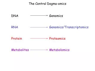
Future Directions in Genomics • Genomics • Is the study of entire genomes • Proteomics • Is the systematic study of all the proteins encoded by a genome • Single nucleotide polymorphisms (SNPs) • Provide useful markers for studying human genetic variation
Concept 20.5: The practical applications of DNA technology affect our lives in many ways • Numerous fields are benefiting from DNA technology and genetic engineering
Medical Applications • One obvious benefit of DNA technology • Is the identification of human genes whose mutation plays a role in genetic diseases