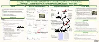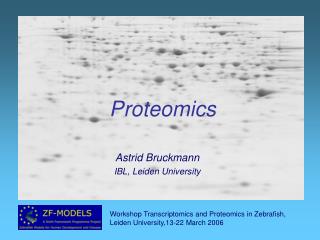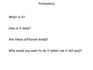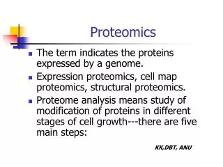
PROTEOMICS LECTURE
PROTEOMICS LECTURE. Genomics. DNA (Gene). Transcription. Transcriptomics. RNA. Translation. Functional Genomics. PROTEIN. Proteomics. Enzymatic reaction. METABOLITE. Metabolomics. The “omics” nomenclature…. Genes Transcripts Proteins Metabolites. Gen Transcript Prote


PROTEOMICS LECTURE
E N D
Presentation Transcript
Genomics DNA (Gene) Transcription Transcriptomics RNA Translation Functional Genomics PROTEIN Proteomics Enzymatic reaction METABOLITE Metabolomics The “omics” nomenclature…
Genes Transcripts Proteins Metabolites Gen Transcript Prote Metabol Sequence of a complete set of ~ome = Gen Prote Genome Proteome ~omics Analysis of the = A few definitions…
Inactive mRNA RNA Degradation control Primary RNA transcript mRNA mRNA DNA Translation control RNA Transport control Modified protein protein RNA Processing control Transcriptional control Post-translational control Why study protein expression? (The steps of gene expression control) (Gygi et al., Mol. Celll. Biol., 1990, p.1720-1730) Cytosol Nucleus
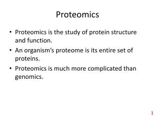
Applications of Proteomics • Mining: identification of proteins (catalog the proteins) • Protein-expression profile: identification of proteins in a particular state of the organism • Protein-network mapping: protein interactions in living systems • Mapping of protein modifications: how and where proteins are modified.
Proteins classes for Analysis • Membrane • Soluble proteins • Nuclear • Chromosome-associated • Phosphorylated • Glycosylated • Complexes
SEPARATION IDENTIFICATION General flow for proteomics analysis
Current Proteomics Technologies • Proteome profiling/separation • 2D SDS PAGE (two-dimensional sodium dodecylsulphate polyacrylamide gel electrophoresis) • 2-D LC/LC (LC = Liquid Chromatography) • 2-D LC/MS (MS= Mass spectrometry) • Protein identification • Peptide mass fingerprint • Tandem Mass Spectrometry (MS/MS) • Quantative proteomics - ICAT (isotope-coded affinity tag)
2D-SDS PAGE gel 1) Sample loading 2) Remove the cover sheet from the IEF gel 3)Place the strip gel in the focusing tray 4) Place the strip on the top of the SDS-PAGE gel
2D-SDS PAGE gel The first dimension (separation by isoelectric focusing) - gel with an immobilised pH gradient - electric current causes charged proteins to move until it reaches the isoelectric point (pH gradient makes the net charge 0)
4 5 Stable pH gradient 6 7 8 9 10 Isoelectric point (pI) • Separation by charge: Low pH: Protein is positively charged At the isolectric point the protein has no net charge and therefore no longer migrates in the electric field. High pH: protein is negatively charged
2D-SDS PAGE gel The first dimension (separation by isoelectric focusing) - gel with an immobilised pH gradient - electric current causes charged proteins to move until it reaches the isoelectric point (pH gradient makes the net charge 0) The second dimension (separation by mass) -pH gel strip is loaded onto a SDS gel -SDS denatures and linearises the protein (to make movement solely dependent on mass, not shape)
Advantages vs. Disadvantages • Not for hydrophobic proteins • Limited by pH range • Not easy for low abundant proteins • Analysis and quantification are difficult • Good resolution of proteins • Detection of posttranslational modifications
2D - LC/LC Peptides all bind to cation exchange column (trypsin) Study protein complexes without gel electrophoresis Successive elution with increasing salt gradients separates peptides by charge Peptides are separated by hydrophobicity on reverse phase column Complex mixture is simplified prior to MS/MS by 2D LC
Reverse Phase column Polypeptides enter the column in the mobile phase… …the hydrophobic “foot” of the polypeptides adsorb to the hydrophobic (non polar) surface of the reverse-phase material (stationary phase) where they remain until… …the organic modifier concentration rises to critical concentration and desorbs the polypeptides
Mass Spectrometry (MS) Stages • Introduce sample to the instrument • Generate ions in the gas phase • Separate ions on the basis of differences in m/z with a mass analyzer • Detect ions
How the protein sequencing works? Ser-Glu-Leu-Ile-Arg-Trp • Use Tandem MS: two mass analyzer in series with a collision cell in between • Collision cell: a region where the ions collide with a gas (He, Ne, Ar) resulting in fragmentation of the ion • Fragmentation of the peptides in the collision cell occur in a predictable fashion, mainly at the peptide bonds (also phosphoester bonds) • The resulting daughter ions have masses that are consistent with known molecular weights of dipeptides, tripeptides, tetrapeptides… Collision Cell Ser-Glu-Leu-Ile-Arg Ser-Glu-Leu-Ile Ser-Glu-Leu Etc…
Tandem Mass Spectrometry Isolates individual peptide fragments for 2nd mass spec – can obtain peptide sequence (trypsin) Compare peptide sequence with protein databases
Advantages vs. Disadvantages • High capital costs • Requires sequence databases for analysis • Determination of MW and aa. Sequence • Detection of posttranslational modifications • High-throughput capability
Protein identification by Peptide Mass fingerprint • Use MS to measure the masses of proteolytic peptide fragments. • Identification is done by matching the measured peptide masses to corresponding peptide masses from protein or nucleotide sequence databases.
(trypsin) eg. MALDI-TOF Compare peptide m/z with protein databases Mass spectometry (MS) Mass spectrometry – method of separating molecules based on mass/charge ratio
Artificially trypsinated Fragmented using trypsin Artificial spectra built Spot removed from gel Protein Identification by MS Spectrum of fragments generated MATCH Library Database of sequences (i.e. SwissProt)
ISOTOPE-CODED AFFINITY TAG (ICAT): a quantitative method • Label protein samples with heavy and light reagent • Reagent contains affinity tag and heavy or light isotopes Chemically reactive group: forms a covalent bond to the protein or peptide Isotope-labeled linker: heavy or light, depending on which isotope is used Affinity tag: enables the protein or peptide bearing an ICAT to be isolated by affinity chromatography in a single step
Example of an ICAT Reagent Biotin Affinity tag: Binds tightly to streptavidin-agarose resin Reactive group: Thiol-reactive group will bind to Cys Linker: Heavy version will have deuteriums at * Light version will have hydrogens at *
100 0 0 600 200 400 550 570 590 How ICAT works? Affinity isolation on streptavidin beads Lyse & Label Quantification MS Identification MS/MS NH2-EACDPLR-COOH Light 100 MIX Heavy Proteolysis (eg trypsin) m/z m/z
Advantages vs. Disadvantages • Yield and non specificity • Slight chromatography differences • Expensive • Tag fragmentation • Meaning of relative quantification information • No presence of cysteine residues or not accessible by ICAT reagent • Estimates relative protein levels between samples with a reasonable level of accuracy (within 10%) • Can be used on complex mixtures of proteins • Cys-specific label reduces sample complexity • Peptides can be sequenced directly if tandem MS-MS is used