Download
1 / 33
470 likes | 1.82k Vues
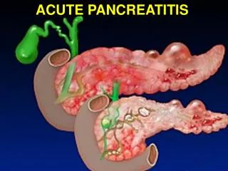
Classification of Acute Pancreatitis. Andrzej D ą browski Department of Gastroenterology and Internal Medicine Medical University of Bialystok, Poland. Why do we need good AP classification?. To improve clinical assessment of the AP

E N D
Classificationof Acute Pancreatitis Andrzej Dąbrowski Department of Gastroenterology and Internal Medicine Medical University of Bialystok, Poland
Why do we need good AP classification? • To improve clinical assessment of the AP • To facilitate communication between treating physicians (uniformity) • For better reporting of clinical studies (common platform for research)
Nicholaes Tulp (1593-1674) of Amsterdam is credited with the first description (1652) of acute pancreatitis. Introduction – historical view Pannala R et al. Pancreas 2009, 38, 355
Introduction – historical view Reginald Fitz in 1889 described 3 forms of acute pancreatitis (hemorrhagic, suppurative, and gangrenous) and proposed that fat necrosis was a sequel of severe pancreatitis Pannala R et al. Pancreas 2009, 38, 355
Introduction – historical view • 1942 – Lagerlof classified pancreatitis as acute and chronic based on clinical, functional, and pathologic observations from autopsy and operative findings. • Beginning in 1955, Joske, then Janowitz in 1957, Howard in 1960, and Dreiling in 1964 developed a comprehensive classification system of pancreatitis based on etiologic factors. • Blumenthal and Probstein in 1959 provided a classification system based on etiology in which they then categorized 163 patients from clinical and autopsy criteria. • 1963 - Marseille Classification of pancreatitis • Acute pancreatitis • Recurretnt acute pancreatitis • Recurrent chronic pancreatitis • Chronic pancreatitis • 1983 – Cambridge classification • 1984 – the second Marseille symposium Frey CF, Pancreas1986, 1, 62
Introduction – historical view • 1983 – Cambridge classification • The Cambridge group defined the severityand complications of acute pancreatitis. • An attackof acute pancreatitis was defined as “mild” if therewas no multisystem failure and “severe” if multisystemfailure occurred and/or there were early orlate, local or systemic complications. • The complicationsidentified for definition were (a) phlegmon,an inflammatory mass in and around the pancreas;(b) pseudocyst, a localized collection of fluid containinghigh concentrations of pancreatic enzymeswithin, adjacent to, or remote from the pancreas:and (c) abscess, pus in or around the pancreas. Frey CF, Pancreas1986, 1, 62
Atlanta classification (1992) Bradley EL III, Arch Surg1993, 128, 586
Atlanta classification (1992) Bradley EL III, Arch Surg1993, 128, 586
Atlanta classification (1992) Bradley EL III, Arch Surg1993, 128, 586
Atlanta classification (1992) Bradley EL III, Arch Surg 1993, 128, 586
Definitions for organ failure and predicted severe AP in guidelines published after 1993 Bollen TL et al., Br J Surg2008, 95, 6
Definitions for organ failure and predicted severe AP in guidelines published after 1993 Bollen TL et al., Br J Surg2008, 95, 6
Definitions of severe AP local complications need revision Contrast-enhanced computed tomography (CT) of a patient with acute pancreatitis 30 days after onset of symptoms. The fluid collection seems to behomogeneous and encapsulated (white arrows) and could be interpreted as a pseudocyst according to the Atlanta Classification. However, at operation the collection was found to contain large amounts of necrotic debris that CT had not shown. Bollen TL et al., Br J Surg2008, 95, 6
CT findings in AP • The interobserver agreement of the Atlanta classification for categorizing peripancreatic collections in acute pancreatitis on CT is poor. The Atlanta classification should not be used to describe complications of acute pancreatitis on CT. Besselink MGH et al, Pancreas2006, 33, 331
CT findings in AP The use of the Atlanta classification on CT in necrotizing pancreatitis. A, Computed tomography scan 12 days after onset of disease. The definitions chosen for this collection were "pseudocyst" (n = 1), "pancreatic abscess" (n = 1), "pancreatic necrosis" (n = 1), and "mixture" (n = 2). B, Computed tomography scan 27 days after onset of disease. The definitions chosen for this collection were "pancreatic abscess" (n = 3) and "mixture" (n = 2). C, Computed tomography scan 31 days after onset of disease. The definitions chosen were "pancreatic necrosis" (n = 1), "pancreatic abscess" (n = 1), "pseudocyst" (n = 1), and "mixture" (n = 2). Besselink MGH et al, Pancreas2006, 33, 331
Need for the revision • Although the Atlanta Classification has proved useful over the following 16 years,many of the definitions proved confusing (usedinconsistently) and have not been accepted or utilized by thepancreatic community (pancreatic gastroenterologists, surgeons, and radiologists).
Revision of the Atlantaclassification of AP • DEFINITION OF ACUTE PANCREATITIS • The clinical definition of AP, whether in the presence or absence of underlying chronic pancreatitis, requires two of the following three features: • 1) abdominal pain suggestive strongly of AP, • 2) serum amylase and/or lipase activity at least 3 times greater than the upper limit of normal, and • 3) characteristic findings of acute pancreatitis on transabdominal ultrasonography or on CECT, which is considered to be the best, most universally available imaging modality. Acute Pancreatitis Classification Working Group. Revision of the Atlantaclassification of acute pancreatitis (3rdrevision) . www. pancreasclub.com/resources/AtlantaClassification.pdf
Revision of the Atlantaclassification of AP • CLINICAL CLASSIFICATION (1st week) • DEFINITION OF SEVERITY OF ACUTE PANCREATITIS • The definition of the severity of acute pancreatitis (during the first week) is based on clinical rather than morphologic parameters (thereafter; over the first week) . • Initially at presentation and over the first 48 hours, patients should be classified temporarily as having severe acute pancreatitis based on the presence of the persistent systemic inflammatory response syndrome (SIRS) and/or developing organ failure. • Several potential risk factors of severity and measurements related to the acute pancreatitis that may reflect severity should be recorded ideally and evaluated prospectively, including age, BMI, hematocrit, APACHE II scores, and serum levels of C-reactive protein. • It should be stressed that serum amylase and lipase activities, while important in the diagnosis of “acute pancreatitis,” are not of any clinical importance in defining the severity of acute pancreatitis. Acute Pancreatitis Classification Working Group. Revision of the Atlantaclassification of acute pancreatitis (3rdrevision) . www. pancreasclub.com/resources/AtlantaClassification.pdf
Revision of the Atlantaclassification of AP • Over the First Week • Over the first week, the distinction between non-severeold term: mild and severe acute pancreatitis depends ultimately on the development of organ failure. • Non-severe acute pancreatitis is defined as the absence of organ failure or the presence of organ failure that does not exceed 48 hours in duration. • The definition of severe acute pancreatitis is the persistence of organ failure that exceeds 48 hours duration (i.e., organ failure recorded at least once during each of three consecutive days). OF>48 hrs Severe AP Acute Pancreatitis Classification Working Group. Revision of the Atlantaclassification of acute pancreatitis (3rdrevision) . www. pancreasclub.com/resources/AtlantaClassification.pdf
Revision of the Atlantaclassification of AP • DEFINITION OF ORGAN FAILURE • Three organ systems should be assessed to define organ failure: respiratory,cardiovascular, and renal. • Organ failure is best and most easily defined in accordancewith the Marshall scoring systemas a score ≥2 for at least one of these threeorgan systems: respiratory (pO2/FIO2); renal (serum creatinine in μmol/l or mg/dl); andcardiovascular (systolic blood pressure in mm Hg). • Multi-system organ failure is defined as two or more organs failing overthe same 2- to 3-day period. Acute Pancreatitis Classification Working Group. Revision of the Atlantaclassification of acute pancreatitis (3rdrevision) . www. pancreasclub.com/resources/AtlantaClassification.pdf
Revision of the Atlantaclassification of AP Marshall Scoring System FIO2 = fraction of inspired oxygen Acute Pancreatitis Classification Working Group. Revision of the Atlantaclassification of acute pancreatitis (3rdrevision) . www. pancreasclub.com/resources/AtlantaClassification.pdf
Revision of the Atlantaclassification of AP • MORPHOLOGIC IMAGING-BASED CLASSIFICATION (used AFTER the first week) • This new classification proposes the use of morphologic CECT criteria to diagnose the specific typeof acute pancreatitis: • Acute interstitial edematous pancreatitis (IEP) • Acute necrotizing pancreatitis. • Presence/absence and site(s) of necrosis, 3 subtypes: • normal pancreatic parenchyma enhancement with peripancreatic fluid collections (fat necrosis) • one or more focal areas of nonenhancing pancreatic parenchyma with peripancreatic fluid collections • without peripancreatic fluid collections • B. Evidence for the presence/absence of infection (FNA, gas within nonenhancing retroperitoneal tissue) Acute Pancreatitis Classification Working Group. Revision of the Atlantaclassification of acute pancreatitis (3rdrevision) . www. pancreasclub.com/resources/AtlantaClassification.pdf
Revision of the Atlantaclassification of AP NECROTIZING PANCREATITIS • Pancreatic Parenchyma: • About 80% of patients with necrotizing pancreatitis have a variable extent of pancreatic parenchymal necrosis on CECT. • The extent of necrosis is quantified in three categories: <30%, 30-50%, and >50% of the total pancreatic parenchyma. • The presence of pancreatic parenchymal non-enhancement differentiates necrotizing pancreatitis from IEP. minimal diffuse gland enlargement on CECT localized Acute Pancreatitis Classification Working Group. Revision of the Atlantaclassification of acute pancreatitis (3rdrevision) . www. pancreasclub.com/resources/AtlantaClassification.pdf
Revision of the Atlantaclassification of AP NECROTIZING PANCREATITIS • Peripancreatic Tissues: • The presence or absence of necrosis in theperipancreatic tissues is more difficult to evaluate by CECT, especially early in thecourse of the disease. • While the presence or absence of necrosis in the peripancreatictissues is not always possible to diagnose definitively with CECT, CECT may suggestthe presence of peripancreatic necrosis by the presence of “thickening” of the paracolicgutters and of the base of the small bowel mesentery, fat stranding and involvement ofthe anterior pararenal spaces, or especially the presence of non-homogeneous fluidcollections containing solid components in one or more areas. Acute Pancreatitis Classification Working Group. Revision of the Atlantaclassification of acute pancreatitis (3rdrevision) . www. pancreasclub.com/resources/AtlantaClassification.pdf
semi-solid necrosis (PNPFC) >4 weeks solid necrosis <1 week liquefaction liquefaction (no resorption) liquefied necrosis (WOPN) • Characteristics of Necrosis: • The relative amount of liquid vs semi-solid components within areas of necrosis varies with the time since onset of necrotizing pancreatitis. • As time evolves, the initially solid necrosis liquefies by a process of liquefaction necrosis. Complete resolution of necrosis (weeks to months later) may occur through liquefaction necrosis and eventual reabsorption of the liquefaction. In some patients, complete reabsorption may never occur. • If resorption does not take place, the area of liquefaction necrosis may persist as an area of walled-off pancreatic necrosis (WOPN; organized necrosis, necroma, or pancreatic sequestration) without symptoms or may cause pain or mechanical obstruction of the duodenum and/or bile duct. Acute Pancreatitis Classification Working Group. Revision of the Atlantaclassification of acute pancreatitis (3rdrevision) . www. pancreasclub.com/resources/AtlantaClassification.pdf
Revision of the Atlantaclassification of AP NECROTIZING PANCREATITIS • Infection: • Depending on the stage of the necrosis (primarily solid, semi-solid, or liquefaction) and the organism(s) involved, the infected necrosis will have varying amounts of suppuration (pus). • In the later stages of infected necrosis, the content may be predominantly pus (in addition to some solid components) as the process of liquefaction necrosis matures. • “Pancreatic abscess” according to the Atlanta Classification in 1992 is a “localized collection of purulent material without significant necrotic material;” most agree that the latter Atlanta definition of “pancreatic abscess” is an exceedingly uncommon finding in necrotizing pancreatitis. The current imaging-based classification does not use the term “pancreatic abscess” in order to avoid this confusion. Acute Pancreatitis Classification Working Group. Revision of the Atlantaclassification of acute pancreatitis (3rdrevision) . www. pancreasclub.com/resources/AtlantaClassification.pdf
Revision of the Atlantaclassification of AP • Both acute IEP and necrotizing pancreatitis can be associated with PANCREATIC AND PERIPANCREATIC FLUID COLLECTIONS Acute Pancreatitis Classification Working Group. Revision of the Atlantaclassification of acute pancreatitis (3rdrevision) . www. pancreasclub.com/resources/AtlantaClassification.pdf
Revision of the Atlantaclassification of AP • ACUTE PERIPANCREATIC FLUID COLLECTIONS (APFCs) (1st 4 weeks after onset of IEP) • a. Sterile • b. Infected Old term: acute fluid collections • These fluid collections arise in patients with IEP, have no solid components, and result from parenchymal and/or peripancreatic inflammation in the absence of necrosis. Resolve spontaneously within 6 weeks 40%, 80% if <6 cm; communication with pancreatic duct seen in 70%. • They exist predominantly adjacent to the pancreas, have no definable wall, and are confined by the normal peripancreatic fascial planes, primarily the anterior pararenal fascia. • APFCs arise presumably from rupture of the main duct or a small peripheral pancreatic ductal side branch or they result from local edema related to the pancreatic inflammation and have no connection with the ductal system. Acute Pancreatitis Classification Working Group. Revision of the Atlantaclassification of acute pancreatitis (3rdrevision) . www. pancreasclub.com/resources/AtlantaClassification.pdf
Revision of the Atlantaclassification of AP • POST-NECROTIC PANCREATIC/PERIPANCREATIC FLUID COLLECTIONS • a. Sterile • b. Infected Old term: acute fluid collections • Fluid collections arising in patients with acute necrotizing pancreatitis are termed PNPFCs to distinguish them from APFCs and pseudocysts. • PNPFCs contain both fluid and necrotic contents to varying degrees. • In PNPFCs, a continuum exists from the initial solid necrosis to liquefaction necrosis, depending on duration of the disease since onset. • PNPFC may or may not have a connection with the pancreatic ductal system. PNPFC (necrosis+fluid) Necrosis WOPN (infected or sterile) late stage Acute Pancreatitis Classification Working Group. Revision of the Atlantaclassification of acute pancreatitis (3rdrevision) . www. pancreasclub.com/resources/AtlantaClassification.pdf
Revision of the Atlantaclassification of AP • PANCREATIC PSEUDOCYST • Non-infected Old term: pancreatic pseudocyst • Pseudocysts on CECT become defined >4 weeks after onset of pancreatitis as a well-circumscribed (clearly evident wall;capsule), usually round or oval, homogeneous fluid collection surrounded by a well-defined wall with no solid necrotic debris within the fluid collection. • Pseudocysts develop from an APFC that persists for >4 weeks after onset of pancreatitis. Prior to 4 weeks, these collections are categorized as APFC. Acute Pancreatitis Classification Working Group. Revision of the Atlantaclassification of acute pancreatitis (3rdrevision) . www. pancreasclub.com/resources/AtlantaClassification.pdf
Revision of the Atlantaclassification of AP • PANCREATIC PSEUDOCYST • Infected (suppurative) Old term: pancreatic abscess • Determination of presence or absence of infection in a pancreatic pseudocyst is also potentially important. • An infected pancreatic pseudocyst contains purulent liquid without an associated solid component (necrosis). • This definition differentiates pseudocyst from infected PNPFC and infected WOPN. As with all peripancreatic fluid collections, image-guided FNA with Gram stain and culture or the presence of extraluminal gas are necessary to confirm the pre-interventional diagnosis of infection. The never-ending process? Acute Pancreatitis Classification Working Group. Revision of the Atlantaclassification of acute pancreatitis (3rdrevision) . www. pancreasclub.com/resources/AtlantaClassification.pdf
Acute pancreatitis has been described for the first time by: Reginald Fitz David Dreiling Nicholaes Tulp Hippokrates of Kos What is the uncommon complication of acute pancreatitis: Respiratory Renal Cardiovascular Gastrointestinal bleeding The true statement about acute peripancreatic fluid collections is: Fluid collections arising in patients with acute necrotizing pancreatitis, but not in patients with acute interstitial edematous pancreatitis. Have no solid components, and result from parenchymal and/or peripancreatic inflammation in the absence of necrosis. Become defined >4 weeks after onset of pancreatitis as a well-circumscribed, usually round or oval, homogeneous fluid collection surrounded by a well-defined wall with no solid necrotic debris within the fluid collection. Contain both fluid and necrotic contents to varying degrees.