Cell
Cell . Cell Theory Plant And Animal Cells Transport. History of the Cell Theory. 1500s-Eyeglass makers-several lenses magnifies objects Anton van Leeuwenhoek: First to describe cells. “ Animalcules” (bacteria) Robert Hooke: Studied cork (dead cells of oak tree); monastery; cells born.

Cell
E N D
Presentation Transcript
Cell Cell Theory Plant And Animal Cells Transport
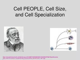
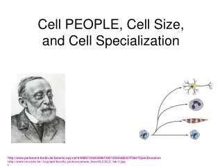
History of the Cell Theory • 1500s-Eyeglass makers-several lenses magnifies objects • Anton van Leeuwenhoek: First to describe cells. “Animalcules” (bacteria) • Robert Hooke: Studied cork (dead cells of oak tree); monastery; cells born. • Robert Brown (1833): Dark structure near the center of the cell (nucleus) • Matthias Schleiden(1838): Plants made of cells • Theodore Schwann (1839): Animals are made of cells. • Rudolf Virchow (1855): Cells come from pre-existing cells
CellTheory • All organisms are composed of one or more cells. • Basic unit of organization of organisms. • All cells come from pre-existing cells.
Modern Cell Theory The cell contains hereditary information which is passed on from cell to cell during cell division. All cells are basically the same in chemical composition and metabolic activities.
Cell Size • 5 to 50 micrometers in diameter • Smallest Mycoplasma bacteria (0.2 micrometers across) • Giant amoebas Chaos chaos (1000 micrometers/1 mm) in diameter; unaided eye
Cell membrane/plasma membrane • Thin flexible barrier • Many cells in direct contact with fluid portion of blood called plamsa.
Nucleus (plural: nuclei) Large membrane enclosed structure that contains genetic material in the form of DNA and controls cell’s activities.
Prokaryotic vs. Eukaryotic PROKARYOTES PRO- “Before” • Generally smaller and simpler (exceptions) • Do not separate genetic material in a nucleus. • All characteristics of life. • Single Cells • Lack organelles • Ex. bacteria EUKARYOTES EU- “True” • Larger, more complex • Dozens of structures • Internal membranes • Highly specialized • Genetic material in nucleus • Single celled, multicellular • Ex. Plants, animals, fungi
Eukaryotic and Prokaryotic Cells Organelles: Membrane bound structures.
Microscopes-use lenses to magify the image of an object by focusing light or electrons
Compound Light Microscope: Uses two or more lenses that lets light throught to magnify objects.Used to examine living cells, small organisms and preserved cells.See cells and structures as small as 1 millionth of a meterMagnification: Up to 1500 x
Compound Microscope • Objective lens-just above specimen-enlarges image • Ocular lens-eyepiece-further magnifies image • Most living things nearly transparent :: use dyes. • Toluidine blue-cell boundaries and nuclei • Fluorescent-give off light of a particular color when viewed under specific wavelengths of light. • Fluorescence microscopy-identify locations of molecules and watch movement.
Electron Microscope: beam of electrons that are focused by a magnetic fieldMagnify object up to 500,000x Produce realistic, 3-D pictures.
Electron Microscopes • Transmission and scanning • Explore cell structures and large protein molecules. • Electrons pass through thin samples-cells and tissues must be cut ultrathin. • Electrons scatter::uses a vacuum • Chemically preserved samples • Nonliving cells and tissues only
Scanning Tunneling Microscope: Probe is brought near specimen. Electrons flow between the tip of the probe and atoms on the specimen’s surface. As probe follows surface contours, 3-D image is created on a computer.Magnification: hundred million times
Transmission Electron Microscope: Aims a beam of electrons through a specimen. Denser objects allow fewer electrons to pass through.Magnification: hundreds of thousands of times
Plasma/Cell Membrane • Structure: A lipid bilayer with protein molecules and carbohydrate chains embedded throughout the bilayer • Function: A selectively permeable membrane which controls what enters and leaves the cell.
Flexible Phospholipids move like water molecules in a current of a lake Fluid Mosaic
Selective Permeability: Process by which the plasma membrane of a cell allows some molecules into the cell while keeping others out.
Phospholipids: Lipids with a phosphate group attached to them.
Phospholipids Glycerol backbone Two fatty acid chains Phospate group
Cholesterol Stabilize phospholipids Prevents fatty acid chains from phospholipids from sticking together
Transport Proteins Allows substances and waste to move through the plasma membrane. Examples: Protein Channels and Carrier Proteins
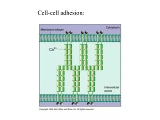
Stick out of cells to help cells identify one another Proteins and Carbohydrates
CellWall • Structure: Fairly rigid structure located outside the plasma membrane. • Function: Shape, support and protection. • Found in plants cells, fungi, most bacteria, and some protists. • Cellulose .
Cilia Structure: Short, numerous, hairlike projections, that move in a wavelike motion. Function: Aid in locomotion and feeding. Flagella Structure: Long projections that move in a whip-like motion. Function: Major means of cell locomotion—unicellular organisms. Cilia And Flagella
Nucleus • Structure: Center of the Cell • Function : Control center of the cell; Contains the direction to make proteins and other important molecules (DNA). • Prokaryotes: DNA in cytoplasm • Plant and animal cells
Chromatin • Structure: Strands of genetic material, DNA; Forms Chromosomes • Function: Master set of directions for making proteins.
Nucleolus • Structure: Prominent structure in the nucleus. • Function: Make Ribosomes (RNA and Proteins)
Nuclear Envelope • Structure :Separates the nucleus from the cytoplasm; Double membrane made of two phospholipid bilayers contain pores for substances to pass through. • Function: Allow materials in and out of nucleus.
Cytoplasm • Structure: Clear, gelatinous fluid inside the cell. Area between cell membrane and nucleus. Dissolved in Cytosol are salts, minerals, and organic compounds. • Function:Contains various cell organelles.
Organelles that Build ProteinsRibosomesEndoplasmic ReticulumGolgi Apparatus
Ribosomes • Structure: Most numerous in cell; no membrane; found free and attached; among smallest of organelles; Made up of RNA and Proteins • Function: Protein Assembly (DNA directions)
Endoplasmic Reticulum • Structure:Complex system of folded membranes suspended in the cytoplasm. • Function: • transportation system between the nucleus and the cytoplasm • Site of chemical reactions • Prepares proteins for export (rER) • synthesizes steroids • regulates calcium levels • breaks down toxic substances (sER) • Smooth (No Ribosomes); Rough (Ribosomes)
Golgi Apparatus/Body • Flattened system of tubular membranes. Flattened stack of pancakes. • Modifies, sorts, and packages proteins and lipids for storage and transport (Cell’s Post Office)
Organelles that Store, Clean-UP, and SupportVacuoles/VesiclesLysosomesCytoskeletonCentrioles
Vacuole Vacule in plants/Vesicle in animals • Structure: Sac surrounded by a membrane. • Function: Stores food, enzymes and other materials needed by the cell, and some vacuoles store waste products.
Plastids • Function: Stores starches and lipids; • Example chloroplast (one type) transfers energy from light to organic compounds.
In plants, plastids may differentiate into several forms, depending upon which function they need to play in the cell. Chloroplasts: for photosynthesis Chromoplasts: for pigment synthesis and storage Amyloplasts: for starch storage Statoliths: for detecting gravity Elaioplasts: for storing fat Proteinoplasts: for storing and modifying protein
Lysosomes • Structure: Small spherical organelles that enclose hydrolytic enzymes within a single membrane. • Function: Digest molecules, old organelles, and foreign substances (engulfs viruses or bacteria)
Cytoskeleton • Structure: Support structure made of tiny rods and filaments. • Function: Form a framework for the cell.
Microtubules and Microfilaments Assist in cell shape and assist organelles in moving from place to place within the cell. Microtubules: Thin hollow cylinders made of proteins. Microfilaments: Thin, solid protein fibers.
Centrioles Pair of cylinder-shaped bodies found in the cells Organize cell division Animal Cells ONLY