Download
1 / 66
660 likes | 811 Vues
Discover the tools and techniques of biotechnology, such as genetic engineering and cloning, used to develop products from living organisms. Learn about DNA manipulation, restriction enzymes, and DNA ligase, with practical applications like PCR.

E N D
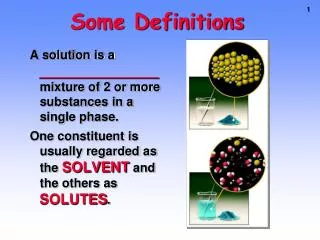
Biotechnology(some definitions) • Biotechnology is the development of products using a living organisms to meet a human need or demand. Note that this includes traditional processes such as wine and cheese production as well as more modern technologies. • Genetic engineering is a technology used to alter the genetic material of living cells in order to make them capable of producing new substances or performing new functions. • Cloning is the production of exact copies (clones) of particular genes or cells.
If you are going to handle genes, first you need to find them (probes), cut them out of a chromosome (restrictionenzymes), glue them (ligation) back into another chromosome (plasmid) then put them into a cell (bacteria, virus, yeast) that can make copies (simple replication or protein synthesis).
Tools of Biotech Collecting the DNA - break open the cells to release DNA - remove unwanted debris -remove unwanted proteins - precipitate out DNA
Restriction Enzymes • Restriction enzymes are proteins produced by bacteria to restrict invasion by foreign DNA (such as viruses). • Restriction enzymes recognise and cut at specific locations along the DNA molecule called recognition sites. • A restriction site is a 4- or 6- base-pair sequence that is a palindrome, ie. The “top” strand read from 5’ to 3’ is the same as the “bottom” strand read from 5’ to 3’. • For example: 5’ GAATTC 3’ 3’ CTTAAG 5’ is the recognition site for the restriction enzymeEcoRI
Restriction Enzyme Action • EcoRI makes one cut between the G and the A in each of the DNA strands. The hydrogen bonds holding the bases together then break. 5’ G AATTC 3’ 3’ CTTAA G 5’ • The single-strands of exposed bases on the cut DNA are called “sticky ends”. Ends cut with the same restriction enzyme can be joined together. • Some restriction enzymes cut the DNA strands directly across from one another producing a “blunt end”. • Hundreds of restriction enzymes have been discovered and are now used.
DNA Ligase • DNA ligase joins together Okazaki fragments on the lagging strand during DNA replication. • Genetic engineers use DNA ligase to join together fragments of DNA (usually from different sources) that have been cut using the same restriction enzyme.
Ligation • Reassembling of DNA strands once they have been cut. e.g. two different pieces of DNA that have been cut with the same restriction enzyme have sticky ends that ‘match’. • These are annealed (bonded) together to get a new piece of DNA in either: Liner or plasmid form • This is then a piece of recombinant DNA. DNA ligase is used to make the pieces join.
PCR – polymerase chain reaction • This is the method by which a small piece of DNA can be quickly copied many times over. It is faster than cloning, you only need a small piece of sample and the sample can be old. • DNA polymerases are the enzymes that copy DNA – to do this they need a template strand and a primer.
PCR(Polymerase Chain Reaction) • A PCR cycle consists of 3 steps: • Separate strands by heating at 98°C for 5 minutes. Allows DNA to unwind. • Cooled and then Addprimers (which are short DNA strands that provide a starting sequence for DNA replication), nucleotides (A, T, G & C) and DNA polymerase. • Incubate, by cooling to 60°C for a few minutes. The primers attach to the single-stranded DNA and DNA polymerase synthesises complementary strands. • Automated DNA sequencing uses thermophilic enzymes so step two is required only for the first cycle. Each cycle takes approx. 5min so many cycles can occur quickly.
PCR animation • AnimationPolymerase Chain Reaction
Applications of PCR • Used by police when have small piece of tissue to identify criminals • Anthropologists and archaeologists to check ancient fossils • Gene checking – i.e. to see if carry cystic fibrosis. • Identify viral genes earlier and quicker than normal methods • Identify genetic disorders in prenatal cells • Detect cancer cells • Identify unknown skeletons
Advantages are: Only need a small piece of tissue Tissue can be old Fast Can be automated Disadvantages; Need to be extremely careful of cross contamination. Pros and cons
Cloning • Cloning can be an entire organism or a single cell many times over. • A vector is any vehicle that carries DNA into a host cell – most modern clones are vectors • Transformation is when external genetic material is assimilated by a cell. • Once engineered DNA needs to be put back in a cell to function.
Natural vectors (application gene cloning) • Plasmids from bacteria are used as vectors These are small rings of DNA separate from the bacteria chromosome. They can easily be removed from the bacteria and cut like other DNA. The two pieces of DNA are joined together and put back into the bacteria.
The bacteria divides and so copies the foreign gene. • These are used in many ways: to make lots of copies of the gene • : or to make bacteria that have a new function • : to make the protein the gene codes for in large quantities.
Other vectors • Viruses: in a virus DNA is a string in a protein coat. New DNA spliced into virus DNA then returned to virus coat. Then infects host cell and replicates (normally bacteria). Some can carry DNA into animals and an advantage is they are normally host specific so only invade certain cells (cystic fibrosis). • Yeast: if protein to be made is too complicated for prokaryote cell then need a eukaryote cell. Yeast is rare as has plasmids so can be used like bacteria.
Other vectors • Can’t always use natural vectors – especially with plant and animal cells. • Electroporation: an electric current is used to force DNA over a cell membrane. • DNA gun: DNA of interest is coated onto microscopic pellets (gold or tungsten) and fired into cells.
Host cells in gene cloning • Usually bacteria as easy to insert genes and replicate quickly. But because prokaryote and eukaryote cells have different enzymes for transcription and translation the prok. does not always read the eukaryote gene correctly, so need to use a eukaryote cell. This is difficult and not many eukaryote cells will take up engineered DNA.
What is DNA cloning? • When DNA is extracted from an organism, all its genes are obtained • In gene (DNA) cloning a particular gene is copied (cloned)
Why Clone DNA? • A particular gene can be isolated and its nucleotide sequence determined • Control sequences of DNA can be identified & analyzed • Protein/enzyme/RNA function can be investigated • Mutations can be identified, e.g. gene defects related to specific diseases • Organisms can be ‘engineered’ for specific purposes, e.g. insulin production, insect resistance, etc.
DNA Synthesis • Scientists can now synthesise short one-sided pieces of DNA, called oligonucleotides. These are made by machines from a computer program. • These oligonucleotides are used as: • Primers for the Polymerase Chain Reaction (PCR).They do this by providing an attachment point for DNA polymerase to synthesise new strands. • Gene probes.These are oligonucleotides that hybridise with specific DNA sequences. A radioactive marker or fluorescent dye is attached to the probe so it is visible.
Separating DNA – Gel Electrophoresis • This method depends on the fact that restriction enzymes produce DNA fragments of different lengths and DNA has a negative charge due to the phosphate groups. • When DNA is exposed to an electrical field, the particles migrate toward the positive electrode • Smaller pieces of DNA can travel further in a given time than larger pieces
Gel Electrophoresis – the method • The gel is made from Agarose - a polysaccharide made from seaweed. Agarose is dissolved in buffer and heated, then cools to a gelatinous solid. • Some gels are made with acrylamide if sharper bands are required
The gel chamber is set up, the ‘comb’ is inserted – this leaves little holes when the gel sets. • The agarose may have a DNA ‘dye’ added (or it may be stained later). The agarose is poured onto the gel block and cooled, then flooded with a buffer solution.
Buffer - the gel slab is submerged (submarine gel) in buffer after hardening • The buffer provides ions in solution to ensure electrical conductivity.
The comb is removed, leaving little ‘wells’ • The DNA samples are mixed with a dense loading dye so they sink into their wells and can be seen
The power source is turned on and the gel is run. The time of the run depends upon the amount of current and % gel, and requires experimentation • At the end of the run the gel is removed (it is actually quite stiff) • The gel is then visualized - UV light causes the bands of DNA to fluoresce
A gel being run Positive electrode Comb Agarose block DNA loaded in wells in the agarose Buffer Black background To make loading wells easier
A gel as seen under UV light - some samples had 2 fragments of DNA, while others had none or one
More…… • Many samples can be run on one gel- but it is important to keep track • Most gels have one lane as a ‘DNA ladder’ - DNA fragments of known size are used for comparison
Still more…. • The DNA band of interest can be cut out of the gel, isolated and purified and then have full biological activity. • Or DNA can be removed from the gel by Southern Blotting
Southern Blotting Summary (Developed by Ed Southern of Edinburgh University). • The method uses gel electrophoresis and hybridisation to find a gene of interest. • Since probes cannot work on a gel, the DNA is transferred to a nylon membrane. • A radioactive probe is then added and hybridises with a specific DNA sequence. • A sheet of photographic film is placed over the membrane and developed to show the position of the probe. • More probes can be used to identify other regions of DNA, since each probe is specific to a particular DNA sequence.
Method • DNA cut with R.E. into small fragments • Separated by gel electrophoresis • Transferred from gel to nylon • Gel soaked to denature DNA • Gel put into long paper towel soaking in salt solution • Nylon membrane placed onto gel, covered in blotting paper and towels
Blotting paper acts as a wick and draws salt solution up through gel • Salt takes DNA with it and transfers it to nylon but in the same position that it was on the gel. • Radioactive probe added that sticks only to the genes of interest and X-ray film can be developed.
DNA Sequencing • This uses gel electrophoresis to find out the order of the nucleotides A, C, T and G on a DNA strand. If you know the order you can then work out the amino acid order of the protein. • Known as the Sanger method after discoverer. • Dideoxynucleotides are used to stop synthesis of a complementary DNA strand at the point they are incorporated. • By using dideoxynucleotide versions of A, C, T and G mixed in with normal versions, it is possible to stop synthesis at every nucleotide.
Since the resulting complementary strands are of different lengths, gel electrophoresis can be used to separate them. • Large modern laboratories use fluorescent dyes and the gels are read by a computer to sequence the DNA.
Why • Understanding a particular DNA sequence can shed light on a genetic condition and offer hope for the eventual development of treatment • DNA technology is also extended to environmental, agricultural and forensic applications
DNA Fingerprinting/profiling • Used to form a genetic fingerprint to identify person, animal or plant. Remember that some of the DNA in humans is common to all organisms, some common to all humans but the unique parts (VNTR and STR) can be used for identification as they are only in one individual.
What is Analyzed in the DNA? • DNA profiling depends on regions of non-coding DNA that show great variability between individuals (are polymorphic which meansmany forms) • Modern profiling uses Short Tandem Repeats, STRs • These are short sequences of DNA, usually 2-5 base pairs (bp) long, that repeat, or ‘stutter’ many times
New Technology • STR analysis has largely replaced the original RFLP analysis (DNA Fingerprinting) developed in 1985 by Dr Alec Jeffreys • RFLP analysis requires good amounts of non-degraded DNA but STR analysis can be done on less than one billionth of a gram (a nanogram) of DNA (as in a single flake of dandruff)
DNA Fingerprinting & DNA Profiling - same or different? • DNA fingerprinting, as developed by Sir Alec Jeffries, produces patterns unique to an individual. It requires good DNA samples and takes 1 - 2 weeks. • DNA profiling produces patterns of inheritance for individual loci, and then uses laws of probability to predict the likelihood of a match. It uses minute amounts of DNA and can be processed within 24 hours
Why Test? • Parentage - e.g. disputes over who is the father of a child & is thus responsible for child support • Determining whether twins are identical or fraternal • Estate cases (these may involve obtaining pathology samples of deceased individuals) • Immigration - establishing that individuals are the true children/parents/siblings in cases of family reunification
Why Test? ctd • Bone marrow transplant monitoring - to check that the transplanted marrow is still present • Determination of maternal cell contamination in chronic villus sampling (used to investigate the possibility that a fetus has a severe inherited disease)- is the tissue sample really fetal? • Etc.
The Steps, II • DNA samples are collected- in the case of parentage testing, from the mother, child and putative (possible) father(s) • They are usually blood, but a buccal (cheek cell) swab is acceptable
The Steps, III • If the samples need transport they must be sent in leak proof containers for the courier’s safety.
The Steps, IV • The samples are processed, and DNA is extracted from each • Primers for each locus are added. Each primer is labeled with a fluorescent marker
The Steps, IV, ctd • DNA Diagnostics currently uses an AmpFlSTR Identifiler TM PCR AmplificationKit which targets 15 STR regions plus a sex specific region. • Kits allow standardization and accuracy, as DNA samples are added to a pre-made mix