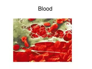
Blood
Blood. 17. Overview of Blood Circulation. Blood leaves the heart via arteries that branch repeatedly until they become capillaries Oxygen (O 2 ) and nutrients diffuse across capillary walls and enter tissues Carbon dioxide (CO 2 ) and wastes move from tissues into the blood.

Blood
E N D
Presentation Transcript
Blood 17
Overview of Blood Circulation • Blood leaves the heart via arteries that branch repeatedly until they become capillaries • Oxygen (O2) and nutrients diffuse across capillary walls and enter tissues • Carbon dioxide (CO2) and wastes move from tissues into the blood
Overview of Blood Circulation • Oxygen-deficient blood leaves the capillaries and flows in veins to the heart • This blood flows to the lungs where it releases CO2 and picks up O2 • The oxygen-rich blood returns to the heart
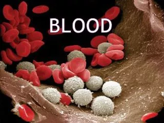
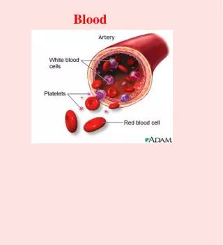
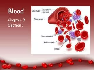
Composition of Blood • Blood is the body’s only fluid tissue • It is composed of liquid plasma and formed elements • Formed elements include: • Erythrocytes, or red blood cells (RBCs) • Leukocytes, or white blood cells (WBCs) • Thrombocytes (Platelets) • Hematocrit – the percentage of RBCs out of the total blood volume
Components of Whole Blood Plasma(55% of whole blood) Buffy coat:leukocyctes and platelets(<1% of whole blood) Formed elements Erythrocytes(45% of whole blood) Withdraw blood and place in tube Centrifuge 1 2 Figure 17.1
Physical Characteristics and Volume • Blood is a sticky, opaque fluid with a metallic taste • Color varies from scarlet (oxygen-rich) to dark red (oxygen-poor) • The pH of blood is 7.35–7.45 • Temperature is 38C, slightly higher than “normal” body temperature • Blood accounts for approximately 8% of body weight • Average volume of blood is 5–6 L for males, and 4–5 L for females
Functions of Blood • Blood performs a number of functions dealing with: • Substance distribution • Regulation of blood levels of particular substances • Body protection
Distribution • Blood transports: • Oxygen from the lungs and nutrients from the digestive tract • Metabolic wastes from cells to the lungs and kidneys for elimination • Hormones from endocrine glands to target organs
Regulation • Blood maintains: • Appropriate body temperature by absorbing and distributing heat • Normal pH in body tissues using buffer systems • Adequate fluid volume in the circulatory system
Protection • Blood prevents blood loss by: • Activating plasma proteins and platelets • Initiating clot formation when a vessel is broken • Blood prevents infection by: • Synthesizing and utilizing antibodies • Activating complement proteins • Activating WBCs to defend the body against foreign invaders
Blood Plasma • Blood plasma contains over 100 solutes, including: • Proteins – albumin, globulins, clotting proteins, and others • Nonprotein nitrogenous substances – lactic acid, urea, creatinine • Organic nutrients – glucose, carbohydrates, amino acids • Electrolytes – sodium, potassium, calcium, chloride, bicarbonate • Respiratory gases – oxygen and carbon dioxide
Formed Elements • Erythrocytes, leukocytes, and platelets make up the formed elements • Only WBCs are complete cells • RBCs have no nuclei or organelles, and platelets are just cell fragments • Most formed elements survive in the bloodstream for only a few days • Most blood cells do not divide but are renewed by cells in bone marrow
Erythrocytes (RBCs) • Biconcave discs, anucleate, essentially no organelles • Filled with hemoglobin (Hb), a protein that functions in gas transport • Contain the plasma membrane protein spectrin and other proteins that: • Give erythrocytes their flexibility • Allow them to change shape as necessary
Erythrocytes (RBCs) Figure 17.3
Erythrocytes (RBCs) • Erythrocytes are an example of the complementarity of structure and function • Structural characteristics contribute to its gas transport function • Biconcave shape that has a huge surface area relative to volume • Discounting water content, erythrocytes are more than 97% hemoglobin • ATP is generated anaerobically, so the erythrocytes do not consume the oxygenthey transport
Erythrocyte Function • Erythrocytes are dedicated to respiratory gas transport • Hemoglobin reversibly binds with oxygen and most oxygen in the blood is bound to hemoglobin • Hemoglobin is composed of the protein globin, made up of two alpha and two beta chains, each bound to a heme group • Each heme group bears an atom of iron, which can bind to one oxygenmolecule • Each hemoglobin molecule can transport four molecules of oxygen
Structure of Hemoglobin Figure 17.4
Hemoglobin • Oxyhemoglobin – hemoglobin bound to oxygen • Oxygen loading takes place in the lungs • Deoxyhemoglobin – hemoglobin after oxygen diffuses into tissues (reduced Hb) • Carbaminohemoglobin – hemoglobin bound to carbon dioxide • Carbon dioxide loading takes place in the tissues
Production of Erythrocytes • Hematopoiesis – blood cell formation • Hematopoiesis occurs in the red bone marrow of the: • Axial skeleton and girdles • Epiphyses of the humerus and femur • Hemocytoblasts give rise to all formed elements
Production of Erythrocytes: Erythropoiesis Figure 17.5
Regulation and Requirements for Erythropoiesis • Circulating erythrocytes – the number remains constant and reflects a balance between RBC production and destruction • Too few red blood cells leads to tissue hypoxia • Too many red blood cells causes undesirable blood viscosity • Erythropoiesis is hormonally controlled and depends on adequate supplies of iron, amino acids, and B12 vitamins
Hormonal Control of Erythropoiesis • Erythropoietin (EPO) release by the kidneys is triggered by: • Hypoxia due to decreased RBCs • Decreased oxygen availability • Increased tissue demand for oxygen • Enhanced erythropoiesis increases the: • RBC count in circulating blood • Oxygen carrying ability of the blood
Erythropoietin Mechanism Imbalance Start Normal blood oxygen levels Stimulus: Hypoxia due to decreased RBC count, decreased availability of O2 to blood, or increased tissue demands for O2 Imbalance Increases O2-carrying ability of blood Reduces O2 levels in blood Erythropoietin stimulates red bone marrow Kidney (and liver to a smaller extent) releases erythropoietin Enhanced erythropoiesis increases RBC count Figure 17.6
Dietary Requirements of Erythropoiesis • Erythropoiesis requires: • Proteins, lipids, and carbohydrates • Iron, vitamin B12, and folic acid • The body stores iron in Hb (65%), the liver, spleen, and bone marrow • Intracellular iron is stored in protein-iron complexes such as ferritin and hemosiderin • Circulating iron is loosely bound to the transport protein transferrin
Fate and Destruction of Erythrocytes • The life span of an erythrocyte is 100–120 days • Old erythrocytes become rigid and fragile, and their hemoglobin begins to degenerate • Dying erythrocytes are engulfed by macrophages • Heme and globin are separated and the iron is salvaged for reuse
Life Cycle of Red Blood Cells Figure 17.7
Erythrocyte Disorders • Anemia – blood has abnormally low oxygen-carrying capacity • It is a symptom rather than a disease itself • Blood oxygen levels cannot support normal metabolism • Signs/symptoms include fatigue, paleness, shortness of breath, and chills
Anemia: Insufficient Erythrocytes • Hemorrhagic anemia – result of acute or chronic loss of blood • Hemolytic anemia – prematurely ruptured erythrocytes • Aplastic anemia – destruction or inhibition of red bone marrow
Anemia: Decreased Hemoglobin Content • Iron-deficiency anemia results from: • A secondary result of hemorrhagic anemia • Inadequate intake of iron-containing foods • Impaired iron absorption • Pernicious anemia results from: • Deficiency of vitamin B12 • Lack of intrinsic factor needed for absorption of B12 • Treatment is intramuscular injection of B12; application of Nascobal (B12 gel)
Anemia: Abnormal Hemoglobin • Thalassemias – absent or faulty globin chain in hemoglobin • Erythrocytes are thin, delicate, and deficient in hemoglobin • Sickle-cell anemia – results from a defective gene coding for an abnormal hemoglobin called hemoglobinS (HbS) • HbS has a single amino acid substitution in the beta chain • This defect causes RBCs to become sickle-shaped in low oxygen situations
Polycythemia • Polycythemia – excess RBCs that increase blood viscosity • Three main polycythemias are: • Polycythemia vera (bone marrow cancer) • Secondary polycythemia (compensation in high attitude areas) • Blood doping
Leukocytes (WBCs) • Leukocytes, the only blood components that are complete cells: • Are less numerous than RBCs • Make up 1% of the total blood volume • Can leave capillaries via diapedesis • Move through tissue spaces • Leukocytosis – WBC count over 11,000 per cubic millimeter (Normal response to bacterial or viral invasion) • Leukopenia WBC abnormally low
Granulocytes • Granulocytes – neutrophils, eosinophils, and basophils • Contain cytoplasmic granules that stain specifically (acidic, basic, or both) with Wright’s stain • Are larger and usually shorter-lived than RBCs • Have lobed nuclei • Are all phagocytic cells
Neutrophils • Neutrophils have two types of granules that: • Take up both acidic and basic dyes • Give the cytoplasm a lilac color • Contain peroxidases, hydrolytic enzymes, and defensins (antibiotic-like proteins) • Neutrophils are our body’s bacteria slayers
Eosinophils • Eosinophils account for 1–4% of WBCs • Have red-staining, bilobed nuclei connected via a broad band of nuclear material • Have red to crimson (acidophilic) large, coarse, lysosome-like granules • Lead the body’s counterattack against parasitic worms • Lessen the severity of allergies by phagocytizing immune complexes
Basophils • Account for 0.5% of WBCs and: • Have U- or S-shaped nuclei with two or three conspicuous constrictions • Are functionally similar to mast cells • Have large, purplish-black (basophilic) granules that contain histamine • Histamine – inflammatory chemical that acts as a vasodilator and attracts other WBCs (antihistamines counter this effect)
Agranulocytes • Agranulocytes – lymphocytes and monocytes: • Lack visible cytoplasmic granules • Are similar structurally, but are functionally distinct and unrelated cell types • Have spherical (lymphocytes) or kidney-shaped (monocytes) nuclei
Lymphocytes • Account for 25% or more of WBCs and: • Have large, dark-purple, circular nuclei with a thin rim of blue cytoplasm • Are found mostly enmeshed in lymphoid tissue (some circulate in the blood) • There are two types of lymphocytes: T cells and B cells • T cells function in the immune response • B cells give rise to plasma cells, which produce antibodies
Monocytes • Monocytes account for 4–8% of leukocytes • They are the largest leukocytes • They have abundant pale-blue cytoplasms • They have purple-staining, U- or kidney-shaped nuclei • They leave the circulation, enter tissue, and differentiate into macrophages
Monocytes • Macrophages: • Are highly mobile and actively phagocytic • Activate lymphocytes to mount an immune response
Summary of Formed Elements Table 17.2
Summary of Formed Elements Table 17.2
Production of Leukocytes • Leukopoiesis is hormonally stimulated by two families of cytokines (hematopoietic factors) – interleukins and colony-stimulating factors (CSFs) • Interleukins are numbered (e.g., IL-1, IL-2), whereas CSFs are named for the WBCs they stimulate (e.g., granulocyte-CSF stimulates granulocytes) • Macrophages and T cells are the most important sources of cytokines • Many hematopoietic hormones are used clinically to stimulate bone marrow
Formation of Leukocytes • All leukocytes originate from hemocytoblasts • Hemocytoblasts differentiate into myeloid stem cells and lymphoid stem cells • Myeloid stem cells become myeloblasts or monoblasts • Lymphoid stem cells become lymphoblasts • Myeloblasts develop into eosinophils, neutrophils, and basophils • Monoblasts develop into monocytes • Lymphoblasts develop into lymphocytes
Formation of Leukocytes Figure 17.11
Leukocytes Disorders: Leukemias • Leukemia refers to cancerous conditions involving white blood cells • Leukemias are named according to the abnormal white blood cells involved • Myelocytic leukemia – involves myeloblasts • Lymphocytic leukemia – involves lymphocytes • Acute leukemia involves blast-type cells and primarily affects children • Chronic leukemia is more prevalent in older people
Leukemia • Immature white blood cells are found in the bloodstream in all leukemias • Bone marrow becomes totally occupied with cancerous leukocytes • The white blood cells produced, though numerous, are not functional • Death is caused by internal hemorrhage and overwhelming infections • Treatments include irradiation, antileukemic drugs, and bone marrow transplants