Nanometer Resolution Imaging of Axonal Transport Defects in Drosophila Alzheimer’s Models
This study investigates axonal transport defects in Drosophila models of Alzheimer’s disease using advanced nanometer resolution detection techniques. Employing fluorescence imaging and multi-peak fitting, we analyze brain segmental nerves to identify and quantify transport abnormalities. The system verification involved the use of fluorescent beads, allowing precise trajectory recovery of APP-YFP vesicles. Our findings utilize a Nikon Eclipse TE2000-U inverted microscope with a 100×/1.40 NA oil objective, supported by a LabVIEW-controlled piezo stage for enhanced pattern recognition and analysis.

Nanometer Resolution Imaging of Axonal Transport Defects in Drosophila Alzheimer’s Models
E N D
Presentation Transcript
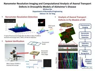
Nanometer Resolution Imaging and Computational Analysis of Axonal Transport Defects in Drosophila Models of Alzheimer’s Disease MinhuaQiu Department of Biomedical Engineering Advisor: Dr. Ge Yang • Analysis of Axonal Transport Defects in Fly Models of AD • Nanometer Resolution Detection Fluorescence Imaging Multi-peak Fitting Brain Segmental Nerves Nanometer Resolution Detection • System Verification Fluorescent beads Trajectory Recovery APP-YFP vesicle imaged A typical Fluorescence Image taken by Nikon Eclipse TE2000-U inverted microscope with a 100×/1.40 NA oil objective Pattern Recognition Nanometer Resolution Piezo Stage Library of Transport Defects Distance: 88 µm Operated by Labview 16 nm Nikon Eclipse TE2000-U inverted microscope 8 nm 0 nm 0 2 4 6 secs